Travis Salzillo1, Yongying Jiang2, Yuri Mackeyev3, Clifton David Fuller1, Caroline Chung1, Seungtaek Choi1, Neil Hughes1, Yao Ding4, Jinzhong Yang4, Sastry Vedam5, Sunil Krishnan3, and Jihong Wang4
1Radiation Oncology, University of Texas MD Anderson Cancer Center, Houston, TX, United States, 2Institute for Applied Cancer Science, University of Texas MD Anderson Cancer Center, Houston, TX, United States, 3Radiation Oncology, Mayo Clinic, Jacksonville, FL, United States, 4Radiation Physics, University of Texas MD Anderson Cancer Center, Houston, TX, United States, 5Radiation Oncology, University of Maryland, Baltimore, MD, United States
1Radiation Oncology, University of Texas MD Anderson Cancer Center, Houston, TX, United States, 2Institute for Applied Cancer Science, University of Texas MD Anderson Cancer Center, Houston, TX, United States, 3Radiation Oncology, Mayo Clinic, Jacksonville, FL, United States, 4Radiation Physics, University of Texas MD Anderson Cancer Center, Houston, TX, United States, 5Radiation Oncology, University of Maryland, Baltimore, MD, United States
There was no visible change in LC-HRMS spectra between the
irradiated samples and unirradiated/reference samples for either contrast agent.
The quantification of the peak areas was uniform among all the samples, and
there was no significant correlation of peak area with dose.
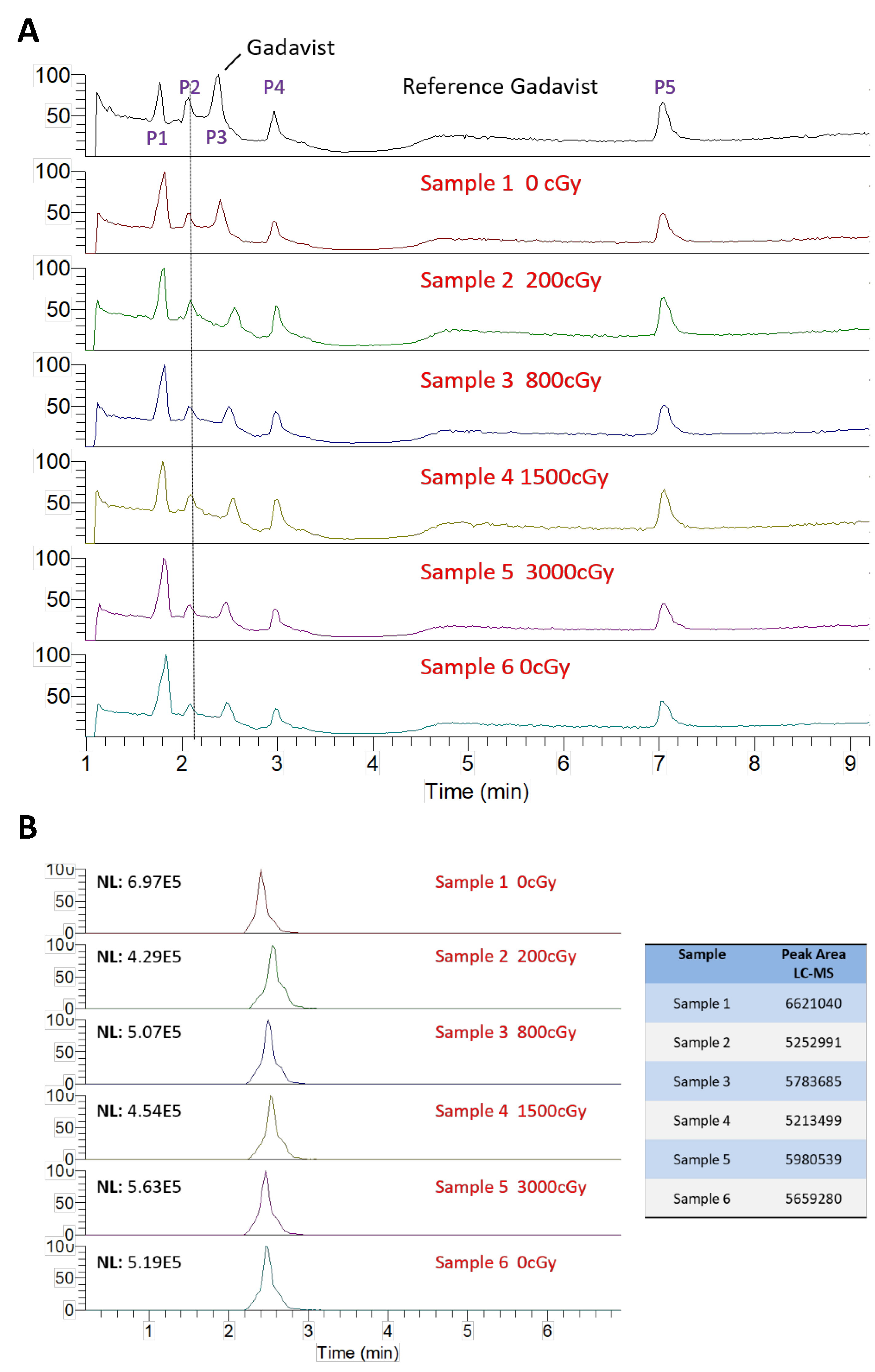
Liquid
chromatography-high-resolution mass spectrometry (LC-HMRS) chromatograms of
gadobutrol samples exposed to various doses of radiation. The entire profile is
displayed in (A) where 5 peaks are visible. Peak P3 was identified as
gadobutrol, which is enlarged and quantified in (B).
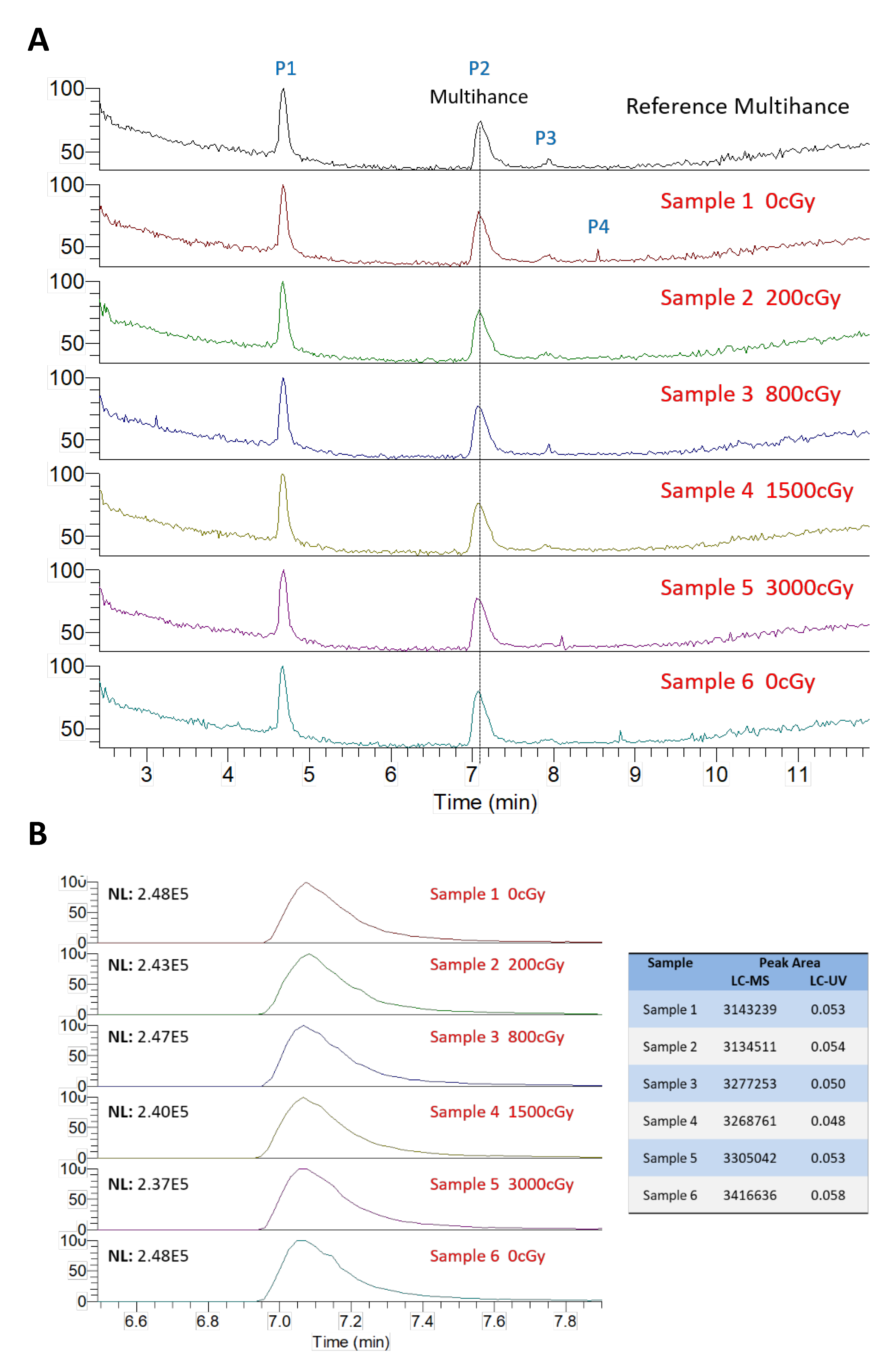
Liquid chromatography-high-resolution
mass spectrometry (LC-HMRS) chromatograms of gadobenate dimeglumine samples exposed to
various doses of radiation. The entire profile is displayed in (A) where
5 peaks are visible. Peak P2 was identified as gadobenate dimeglumine, which is enlarged and
quantified in (B).