Iichiro Osawa1, Eito Kozawa1, Yuya Yamamoto1, Sayuri Tanaka1, Taira Shiratori1, Akane Kaizu1, Kaiji Inoue1, and Mamoru Niitsu1
1Saitama Medical University Hospital, Saitama, Japan
1Saitama Medical University Hospital, Saitama, Japan
The infundibular
recess (IR) was enhanced on HT2-FLAIR after an intravenous gadolinium
injection. Enhancement was stronger on post-contrast images than on 4-h delayed
post-contrast images. IR showed stronger enhancement than other CSF spaces.
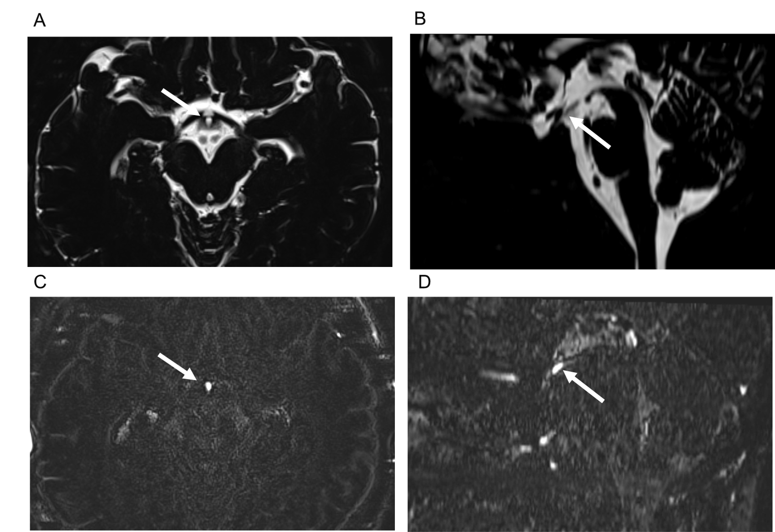
Figure 1. Contrast enhancement of the infundibular recess.
The infundibular recess is hyperintense on axial (A, arrow) and midsagittal
reformatted (B, arrow) MR cisternography (MRC). It shows contrast enhancement
on axial (C, arrow) and midsagittal reformatted (D, arrow) post-contrast HT2-FLAIR.
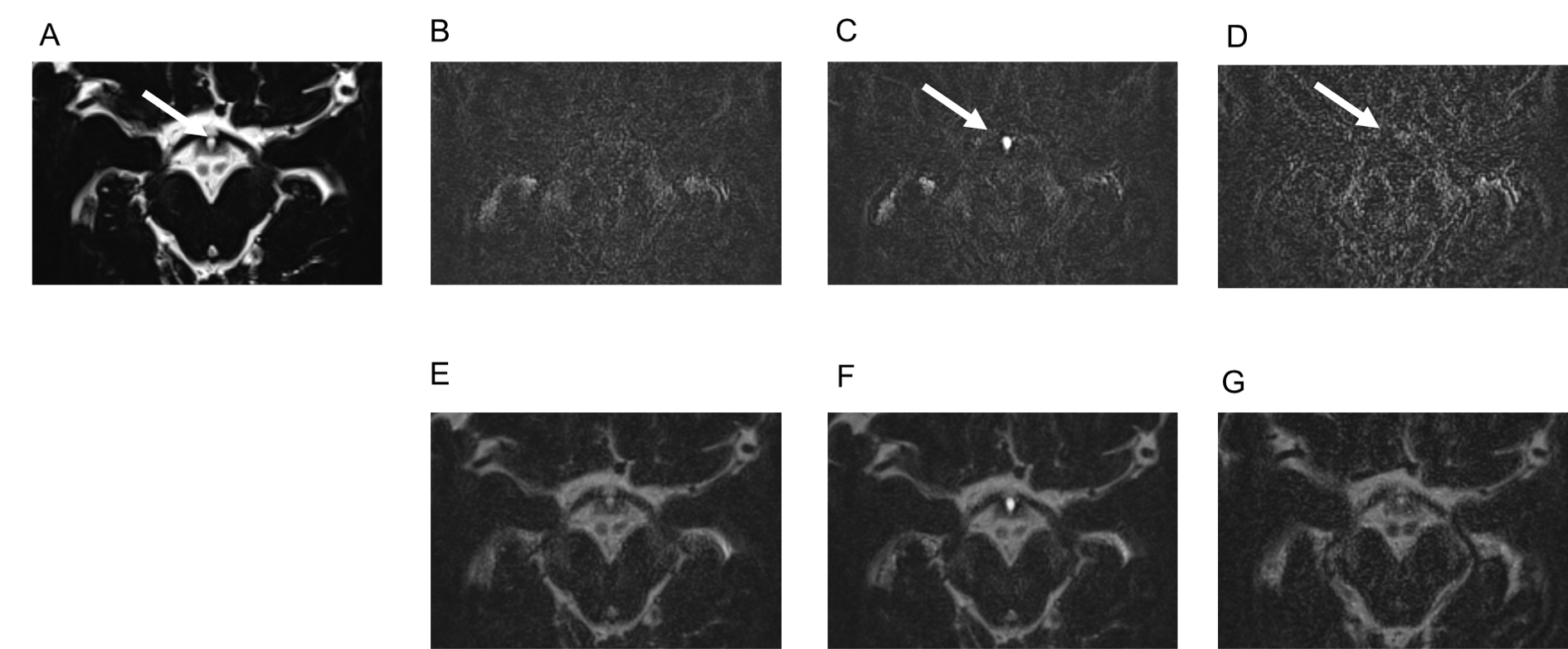
Figure 2. Chronological changes in contrast enhancement in the
infundibular recess.
The infundibular recess (IR) is hyperintense on MR cisternography (MRC)
(A, arrow). On HT2-FLAIR, compared with a pre-contrast image (B), a post-contrast
image (C, arrow) of IR shows stronger enhancement. A 4-h delayed post-contrast
image (D, arrow) shows weaker enhancement. We fuse MRC and each HT2-FLAIR into
one image (E, F, and G), and enhancement on post-contrast HT2-FLAIR (F)
corresponds to IR hyperintensity on MRC.