Patrick Metze1, Hao Li2, and Volker Rasche1,2
1Internal Medicine II, Ulm University Medical Center, Ulm, Germany, 2Core Facility Small Animal Imaging (CF-SANI), Ulm University, Ulm, Germany
1Internal Medicine II, Ulm University Medical Center, Ulm, Germany, 2Core Facility Small Animal Imaging (CF-SANI), Ulm University, Ulm, Germany
Radial
tiny golden angle MRI allows for reconstruction of high-quality respiratory and
cardiac gated images and sliding-window real-time imaging from the same
continuous acquisition, enabling e.g. simultaneous perfusion and function
assessment.
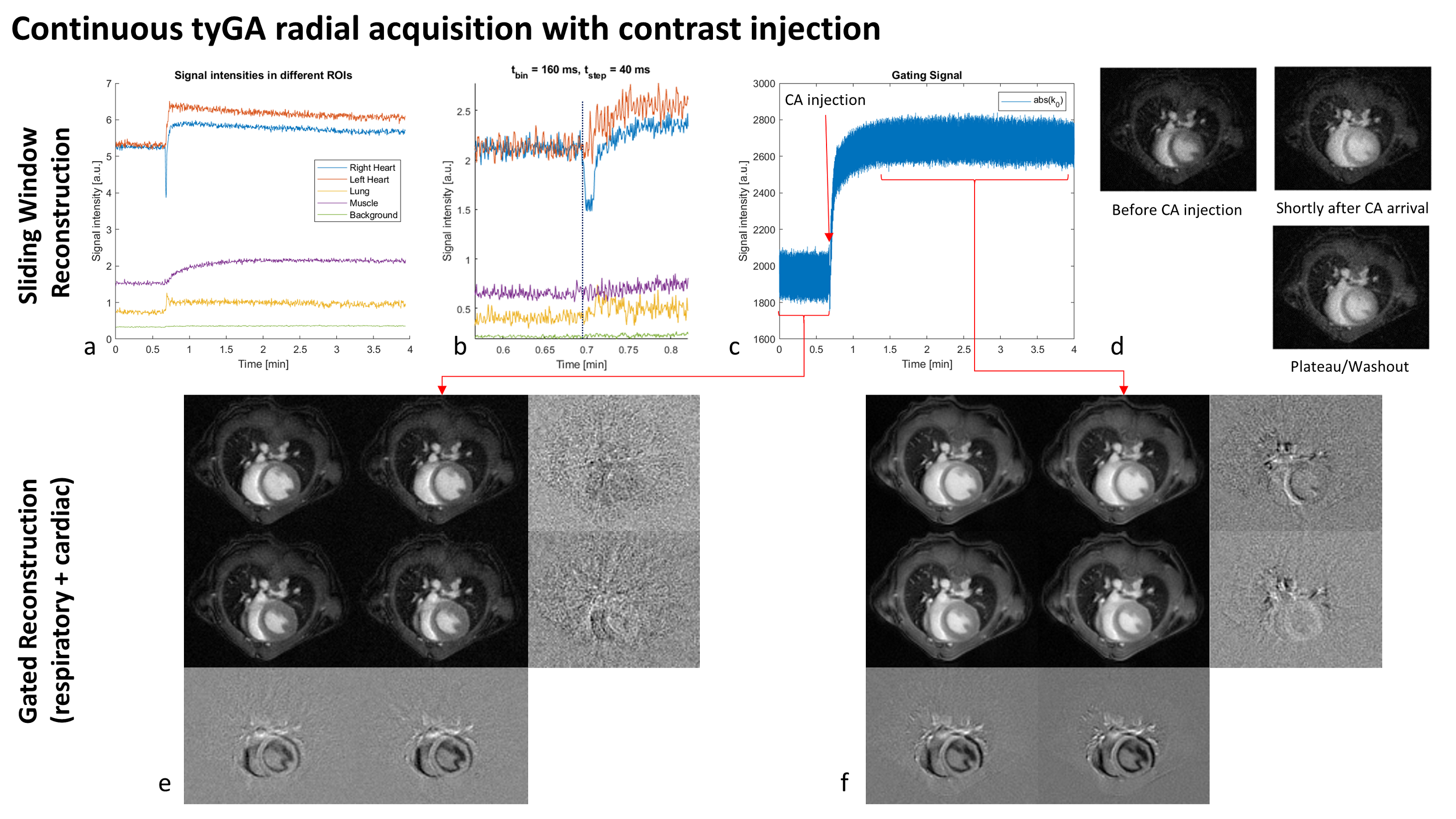
Figure 4: Continuous
tyGA acquisition with simultaneous administration of contrast agent. a) and c)
show image ROI and k-space intensities over the whole acquisition, b) shows the
image intensities around the timepoint of administration. d) shows example
sliding-window images and e) and f) correspond to dual gated reconstructions
before and after contrast administration. Row-wise differences indicate
different respiration stages, column-wise differences show the influence of the
cardiac phase.
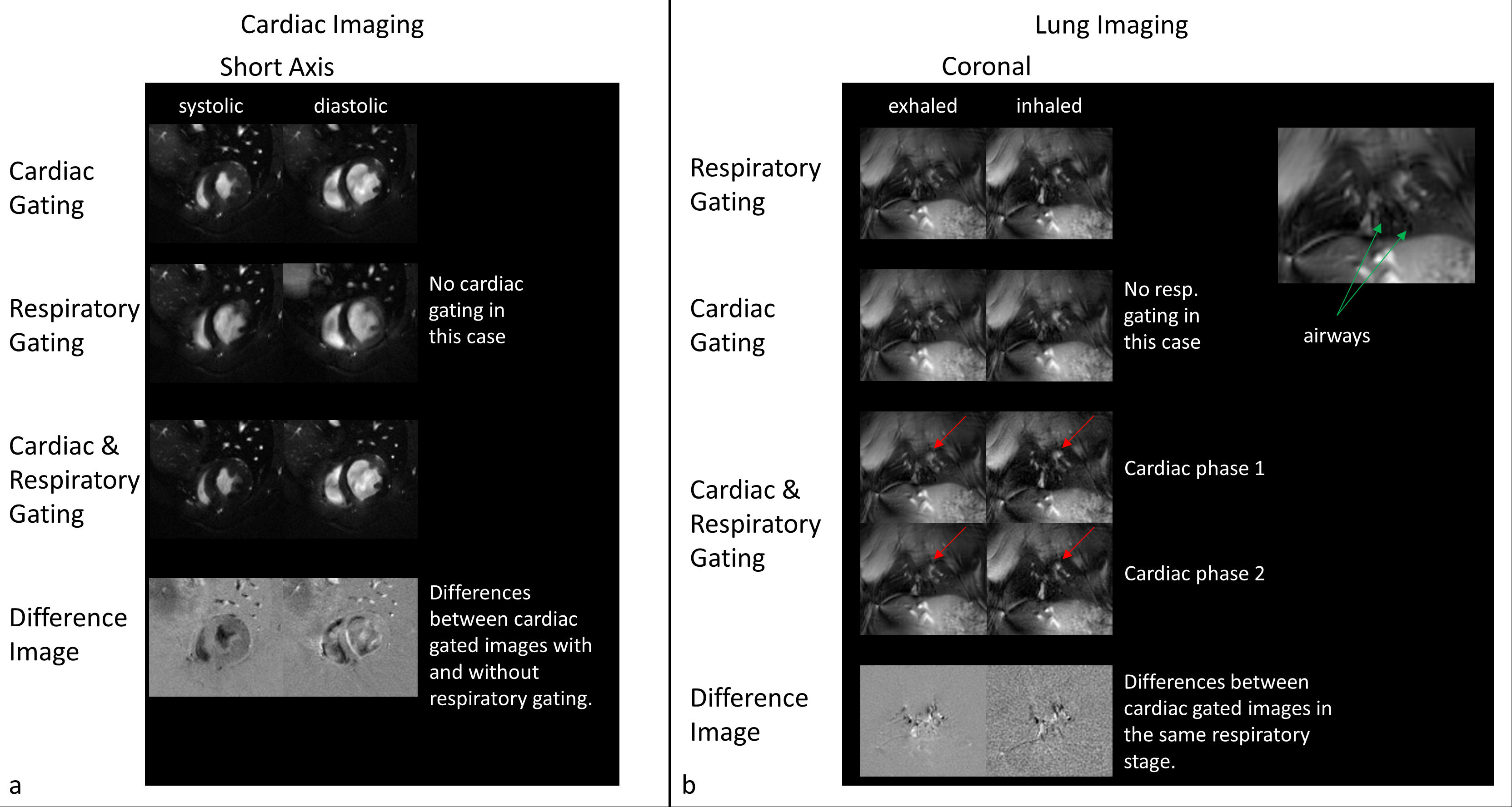
Figure 3: Self-gated
reconstructions of an exemplary cardiac (SAx) and lung (coronal) acquisition. Due to the properties of the tyGA trajectory, artefact levels for respiratory ungated cardiac
imaging is very low. However, differences to respiratory gated imaging can not only be seen
in the vessels, but also in the heart itself. Cardiac
gating seems mandatory for lung imaging, as shape and position of vessels
clearly change with the heartbeat and could influence functional measurements.