Aurea B Martins-Bach1, Mohamed Tachrount1, Cristiana Tisca1, Lily Qiu1, Shoshana Spring2, Jacob Ellegood2, Brian J Nieman2, John G. Sled2, Remya Raghavan-Nair 3, Elizabeth Fisher4, Thomas Cunningham3, Jason Lerch1, and Karla Miller1
1Wellcome Centre for Integrative Neuroimaging, University of Oxford, Oxford, United Kingdom, 2Mouse Imaging Centre, The Hospital for Sick Children, Toronto, ON, Canada, 3Mammalian Genetics Unit, MRC Harwell Institute, Oxford, United Kingdom, 4Department of Neuromuscular Diseases, Institute of Neurology, University College London, London, United Kingdom
1Wellcome Centre for Integrative Neuroimaging, University of Oxford, Oxford, United Kingdom, 2Mouse Imaging Centre, The Hospital for Sick Children, Toronto, ON, Canada, 3Mammalian Genetics Unit, MRC Harwell Institute, Oxford, United Kingdom, 4Department of Neuromuscular Diseases, Institute of Neurology, University College London, London, United Kingdom
ALS is a devastating disease characterized by aggregates of TDP-43 protein in the brain. We assessed structural and microstructural alterations in the brain of TDP-M323K mice with preclinical MRI. This mouse model of ALS presented imaging alterations compatible with neurodegeneration.
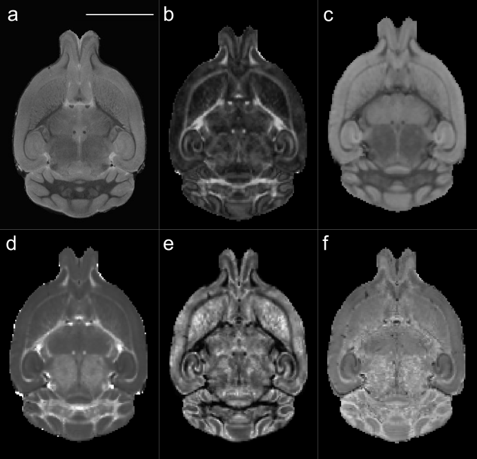
Figure 1. Representative images and diffusion MRI maps. (a) T2w structural image for volumetric
analysis. b-d: Parametric maps extracted from diffusion MRI after modelling the
signal to the diffusion kurtosis model: FA: fractional anisotropy (b), MD: mean
diffusivity (c), and MK: mean kurtosis (d). e,f: Parametric maps extracted using
the NODDI model: OD: orientation dispersion (e) and ICVF: intracellular volume
fraction (f). Scale bar: 5 mm.
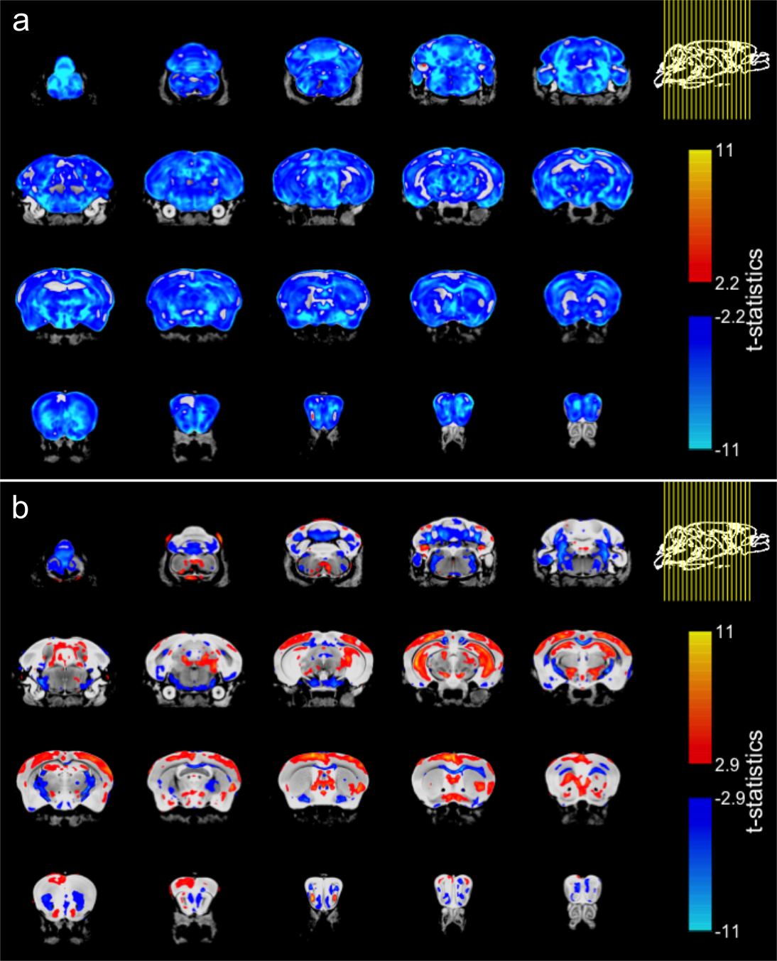
Figure 3. Effect of genotype in brain volume in TDP-M323K mice. (a) When compared directly to WT mice, TDP-M323K mice showed smaller volumes in almost all regions of the brain. (b) When volumes relative to the whole brain size were compared, TDP-M323K mice showed atrophy mainly in the white matter, while regions in the cortex and hippocampus had relatively increased volume. Most of the spatial patterns in t-statistics maps are bilaterally symmetric. Atrophic regions in TDP-M323K mice are show in blue and hypertrophic regions are show in red/yellow. False discovery rate of 5%.