Rohit Mahar1, Huadong Zeng2, Anthony Giacalone3, Mukundan Ragavan3, Thomas H. Mareci3, and Matthew E. Merritt3
1Biochemistry and Molecular Biology, University of Florida, Gainesville, FL, United States, 2Advanced Magnetic Resonance Imaging and Spectroscopy (AMRIS) Facility, University of Florida, Gainesville, FL, United States, 3Department of Biochemistry and Molecular Biology, University of Florida, Gainesville, FL, United States
1Biochemistry and Molecular Biology, University of Florida, Gainesville, FL, United States, 2Advanced Magnetic Resonance Imaging and Spectroscopy (AMRIS) Facility, University of Florida, Gainesville, FL, United States, 3Department of Biochemistry and Molecular Biology, University of Florida, Gainesville, FL, United States
The
gradient echo methods demonstrate the first efficacious use of HDO generated
from [2H7]glucose as a means for producing metabolically
sensitive MR images. The HDO image should serve as an accurate biomarker of
glycolytic flux and recapitulate glx kinetics in the functioning brain.
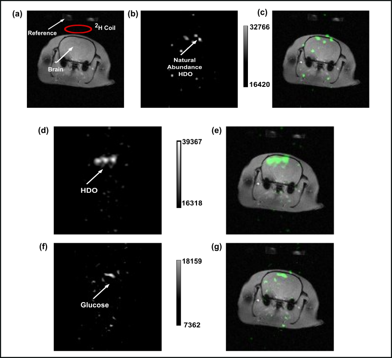
Figure 2:
Multi-gradient echo (MGE) imaging to separate the contribution of [2H7]glucose
and HDO to DMRI image of the rat brain. Image
(d) represents the deuterium image of HDO-only and (e) is the overlaid images
of (a) proton (grey) and (d) deuterium (green) images due to HDO-only in the
rat brain.
Image
(f) represents the deuterium image from the contribution of [2H7]glucose-only
and image (g) is the overlaid image of (a) proton and (f) deuterium [2H7]glucose-only
images.
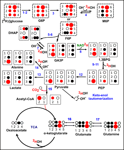
Figure 3: Schematic
representation of the production of deuterated lactate, glutamate/glutamine
(glx), and HDO from [2H7]glucose during glycolysis and
the TCA cycle. Deuterium (2H) loss has been shown in the form of 2HOH,
and NAD2H. Small and large red filled circles represent 1 and 2
deuterium atoms respectively, black filled, and empty circles represent
hydrogen atoms and quaternary carbons, respectively.