Natasha N Knier1,2 and Paula J Foster1,2
1University of Western Ontario, London, ON, Canada, 2Robarts Research Institute, London, ON, Canada
1University of Western Ontario, London, ON, Canada, 2Robarts Research Institute, London, ON, Canada
In this study, we demonstrate that brain metastatic breast cancer cells can be labeled with Synomag-D™ and detected in vivo in the mouse brain with MPI. Iron content within was quantified, addressing a major limitation of iron-based MRI cell tracking.
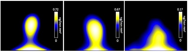
Figure 3: MPI of mouse brains injected with 2x10⁵ Synomag-D
labeled 231BR cells at day 0, imaged with a FOV of 6 cm x 4
cm.
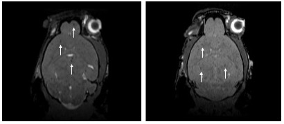
Figure 4: Representative MRI
with a FOV of 1.5 cm x 1.5 cm of mouse brains injected with 2.5 x 10⁵ 231BR
cells labeled with MPIO at Day 0. Discrete signal voids representing iron
labeled cancer cells appear throughout the brain (white arrows).