Julian Mevenkamp1, Yvonne M.H. Bruls1,2, Robin A. de Graaf3, Joachim E. Wildberger1, Matthijs K.C. Hesselink2, Lucas Lindeboom1,2, and Vera B. Schrauwen-Hinderling1,2
1Department of Radiology & Nuclear Medicine, Maastricht University Medical Center, Maastricht, Netherlands, 2Department of Nutrition & Movement Sciences, Maastricht University, Maastricht, Netherlands, 3Department of Radiology & Biomedical Imaging, Yale School of Medicine, New Haven, CT, United States
1Department of Radiology & Nuclear Medicine, Maastricht University Medical Center, Maastricht, Netherlands, 2Department of Nutrition & Movement Sciences, Maastricht University, Maastricht, Netherlands, 3Department of Radiology & Biomedical Imaging, Yale School of Medicine, New Haven, CT, United States
Our newly developed homonuclear BIRD filter suppresses α-ATP resonances in 31P MRS and
therefore allows the quantification of NADH and NAD+ resonances on
clinical scanners at 3T. However, separation of NADH and NAD+
resonances remains challenging.
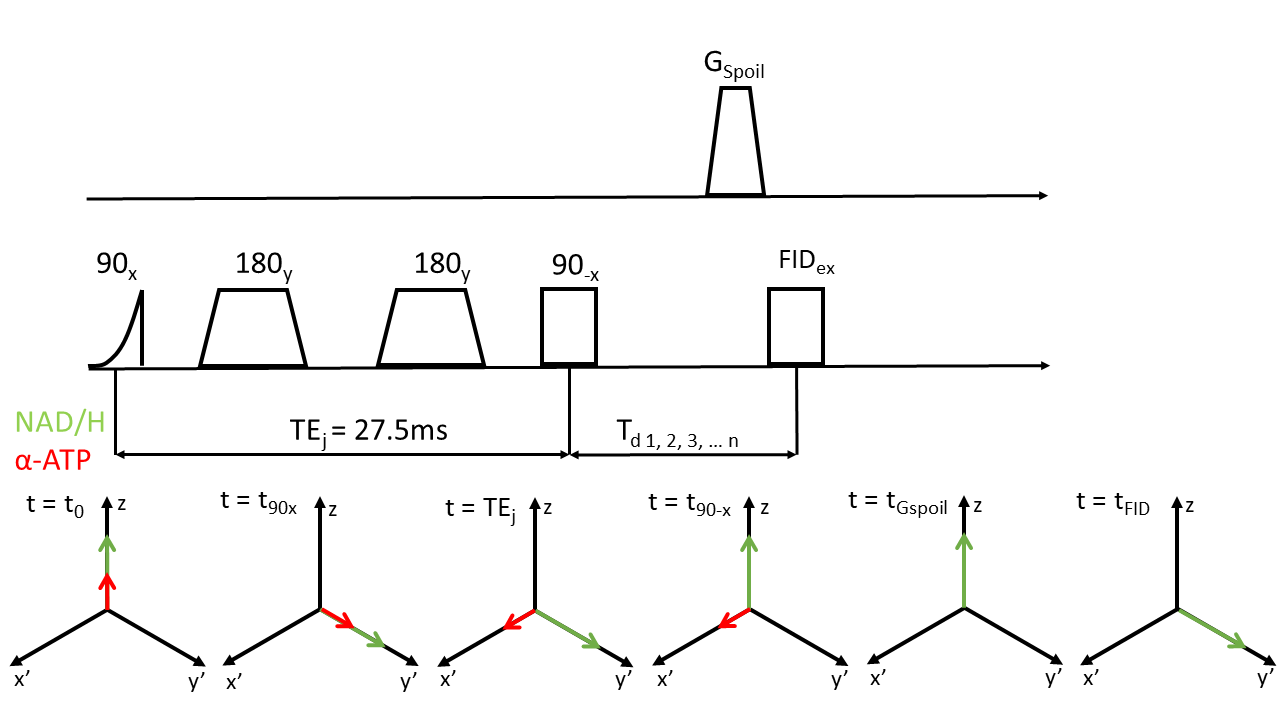
Figure 1: Schematics of
homonuclear BIRD (HB) filter. After an initial adiabatic 90⁰ hard pulse and two
adiabatic 180⁰ pulses, α-ATP spins acquire a phase of 90⁰ with
respect to uncoupled spins at t=TEj =1/2J and end up aligned with y’ (t=TEj).
At t90-x a non-selective 90⁰ block pulse flips spins with J-coupling
constants different from that of α-ATP about the y’-axis back towards the
z-axis. Spins remaining spins in the x’-y’ plane are then dephased by GSpoil. Zero quantum coherences (ZQC) are
removed by co-adding signals acquired with a variable delay Td.
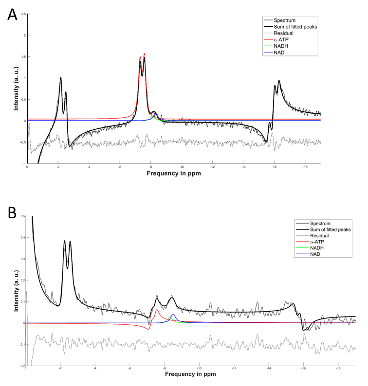
Figure 3: Comparison between NAD+/H fitting results from (A)
FID and (B) HB filtered spectra. NAD+/H resonances are clearly more
separated from α-ATP in HB filtered spectra than in the FID.