Lucas Narciso1,2, Tracy Ssali1,2, Linshan Liu1, Heather Biernaski1, John Butler1, Laura Morrison1, Jennifer Hadway1, Jeffrey Corsaut1, Justin W. Hicks1, Michael C. Langham3, Felix W. Wehrli3, Hidehiro Iida4, and Keith St Lawrence1,2
1Lawson Health Research Institute, London, ON, Canada, 2Department of Medical Biophysics, Western University, London, ON, Canada, 3University of Pennsylvania Perelman School of Medicine, Philadelphia, PA, United States, 4University of Turku and Turku PET Centre, Turku, Finland
1Lawson Health Research Institute, London, ON, Canada, 2Department of Medical Biophysics, Western University, London, ON, Canada, 3University of Pennsylvania Perelman School of Medicine, Philadelphia, PA, United States, 4University of Turku and Turku PET Centre, Turku, Finland
Whole-brain cerebral
blood flow, oxygen extraction fraction, and cerebral metabolic rate of oxygen
measurements from OxFlow-MRI and PET were in good agreement. OxFlow was
sensitive to reduced metabolism due to increased anesthetics.
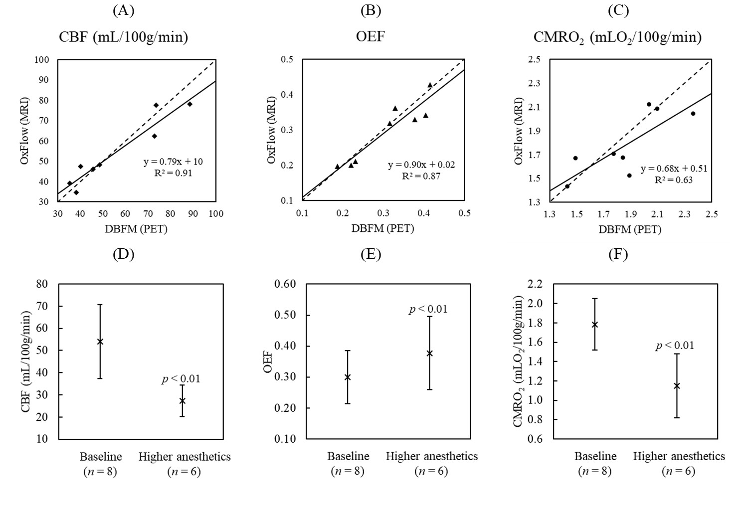
Figure
2. Comparison between (A) CBF, (B) OEF and (C) CMRO2
estimates from DBFM (PET-only technique) and OxFlow (n = 8). No significant difference was observed for all
three measurements. The dashed and solid lines represent the identify and
regression lines, respectively. A significant anesthetics-induced reduction in
(D) WB CBF (27.3 ± 7.0 mL/100g/min) was accompanied by an
increase in (E) WB OEF (0.38 ± 0.12), resulting in a significant
decrease in (F) CMRO2 (1.15 ± 0.33 mLO2/100g/min).
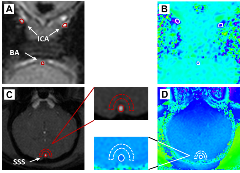
Figure
1. Magnitude and phase images from the slices used
to estimate (A)-(B) WB CBF and (C)-(D) SvO2. The regions-of-interest
(red dashed) were transferred from the magnitude to the phase image (in white).
Images are from one representative animal.