Duen-Pang Kuo1,2, Yi-Tien Li1,3, Chen-Yin Ou1, Yung-Chieh Chen1,2, and Cheng-Yu Chen1,2
1Translational Imaging Research Center, Taipei Medical University Hospital, Taipei, Taiwan, 2Department of Medical Imaging, Taipei Medical University Hospital, Taipei, Taiwan, 3Neuroscience Research Center, Taipei Medical University, Taipei, Taiwan
1Translational Imaging Research Center, Taipei Medical University Hospital, Taipei, Taiwan, 2Department of Medical Imaging, Taipei Medical University Hospital, Taipei, Taiwan, 3Neuroscience Research Center, Taipei Medical University, Taipei, Taiwan
Significant DTI changes were found at the boundary of bilateral thalami where high shear stress was expected, suggesting that disinhibition of inhibitory circuits from the thalamic reticular nucleus may play a role in thalamocortical
dysrhythmia.
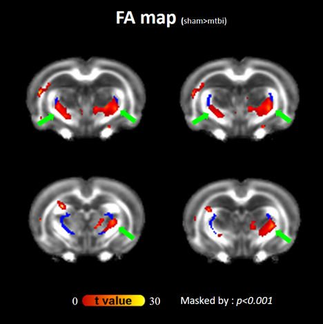
Figure 1 Statistical
parametric maps of the FA changes between pre- and post-impact Significantly increases in
FA at bilateral thalamic borders (green arrows) were observed post-impact as compared
with pre-impact. The dark blue indicated the location of bilateral TRN. These
maps were further masked by the threshold of group-averaged FA > 0.15.
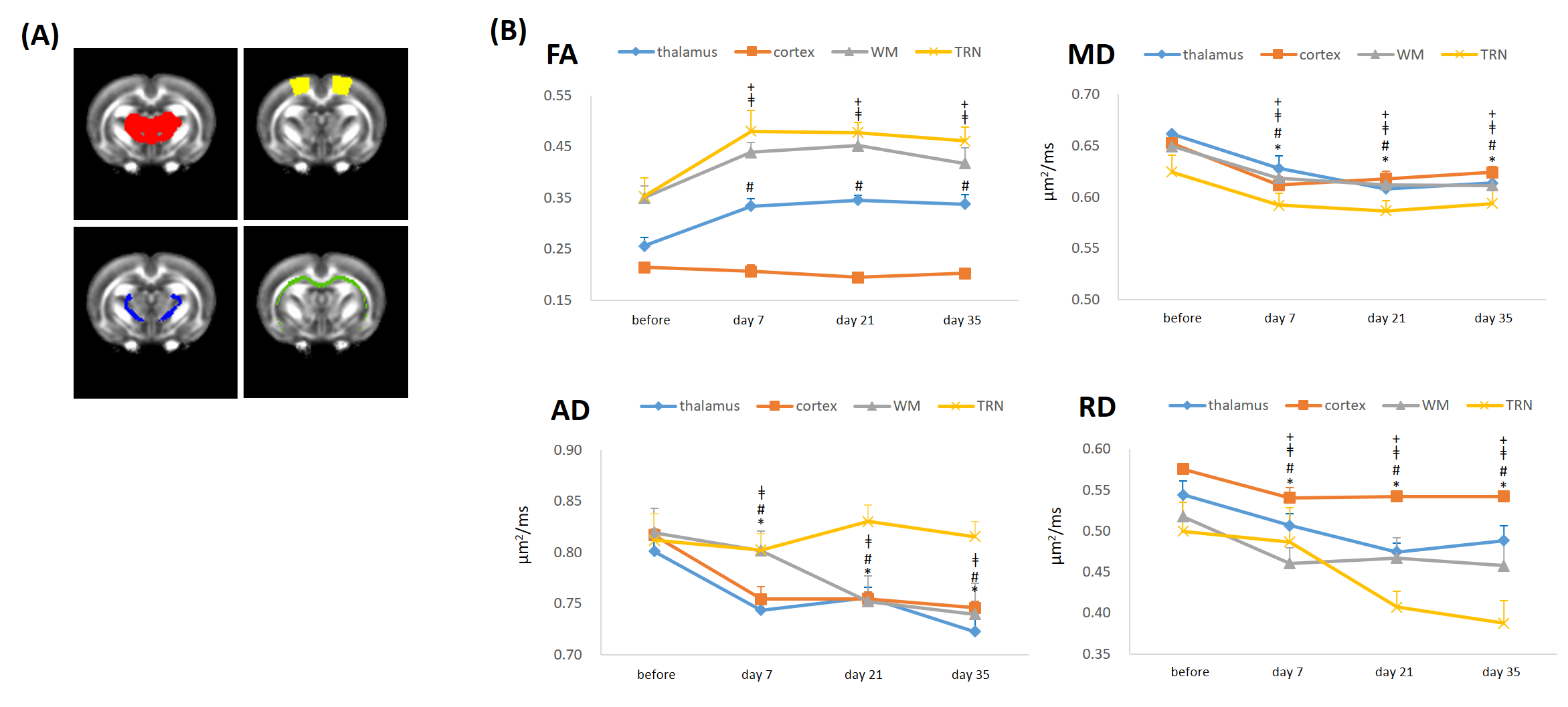
Figure 3. Longitudinal changes in DTI
metrics after impact (A)
Bilateral segmented ROIs showing the areas of thalamus (red),cortex (yellow),
TRN(blue) and WM(green) (B) Longitudinal follow-up during the first 35 days
shows that FA increased in thalamus, WM and TRN at day 7 as well as during the
follow-up. Contrary to FA, MD, AD and RD decreased with time except for AD
in TRN. (thalamus comparison with baseline[before impact]: # P <0.01;
cortex comparison with baseline: * P <0.01; WM comparison with
baseline: ǂ P <0.05; TRN comparison with baseline: + P <0.05 ; data presented as mean ± sd)