Cristiana Tisca1, Mohamed Tachrount 1, Frederik Lange1, Chaoyue Wang1, Lily Qiu1, Javier Barallobre-Barreiro2, Marika Fava2, Manuel Mayr2, Jason Lerch1,3,4, Aurea Martins-Bach1, and Karla Miller1
1Wellcome Centre for Integrative Neuroimaging, FMRIB, Nuffield Department of Clinical Neurosciences, University of Oxford, Oxford, United Kingdom, 2British Heart Foundation Centre of Research Excellence, King's College London, London, United Kingdom, 3Mouse Imaging Centre, Hospital for Sick Children, Toronto, ON, Canada, 4Department of Medical Biophysics, University of Toronto, Toronto, ON, Canada
1Wellcome Centre for Integrative Neuroimaging, FMRIB, Nuffield Department of Clinical Neurosciences, University of Oxford, Oxford, United Kingdom, 2British Heart Foundation Centre of Research Excellence, King's College London, London, United Kingdom, 3Mouse Imaging Centre, Hospital for Sick Children, Toronto, ON, Canada, 4Department of Medical Biophysics, University of Toronto, Toronto, ON, Canada
We show that male Vcan mice display related dMRI phenotypes to those associated with mutations in VCAN in humans. This gives us the ability to investigate the biological interpretations of human genome-wide association studies results using histology.
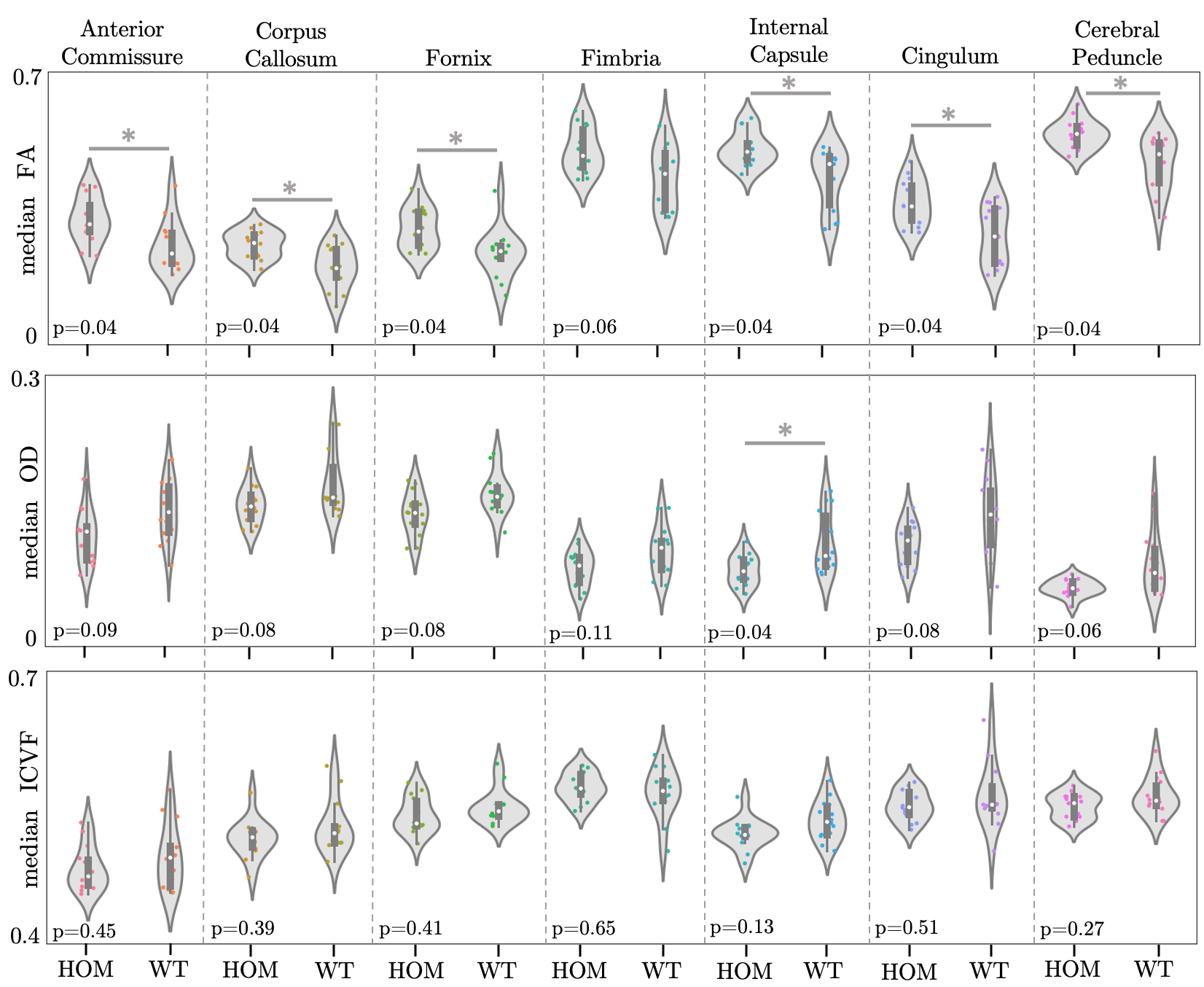
Figure 3. Violin plots of median FA, OD and ICVF values within 7 ROIs for male mice. *: p-value ≤0.05, FDR-corrected (5%).
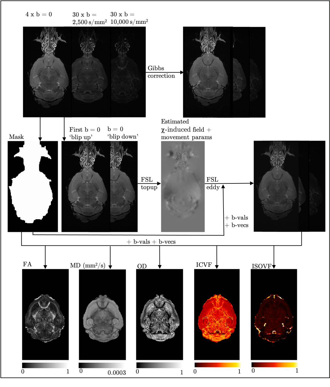
Figure 1. Illustration of the dMRI pipeline. All 64 volumes are corrected for Gibbs ringing6. The data was first corrected for susceptibility-induced off-resonance field distortions and movement using topup7 and eddy8. The diffusion tensor model (FSL DTIFIT) and the NODDI12 model (FSL cuDIMOT13 implementation; d||=5.6x10⁻⁴, diso=1x10⁻³mm2/s, dot compartment included) were fit to the data to produce FA, MD, OD, ICVF and isotropic volume fraction (ISOVF) maps.