Jabrane Karkouri1, Stanislav Frištyk2, Lucian AB Purvis3, Christopher T. Rodgers*1, and Ladislav Valkovic*3
1Wolfson Brain Imaging Center, University of Cambridge, Cambridge, United Kingdom, 2Department of Electromagnetic and Biomedical Engineering, University of Žilina, Zilina, Slovakia, 3Oxford Centre for Clinical Magnetic Resonance Research, University of Oxford, Oxford, United Kingdom
1Wolfson Brain Imaging Center, University of Cambridge, Cambridge, United Kingdom, 2Department of Electromagnetic and Biomedical Engineering, University of Žilina, Zilina, Slovakia, 3Oxford Centre for Clinical Magnetic Resonance Research, University of Oxford, Oxford, United Kingdom
Phosphorus
magnetic resonance spectroscopy delivers unique information to aid
our understanding of cardiac metabolism. In this study, we investigate the
feasibility of absolute concentration of phosphorus metabolites in the human
heart at 7T.
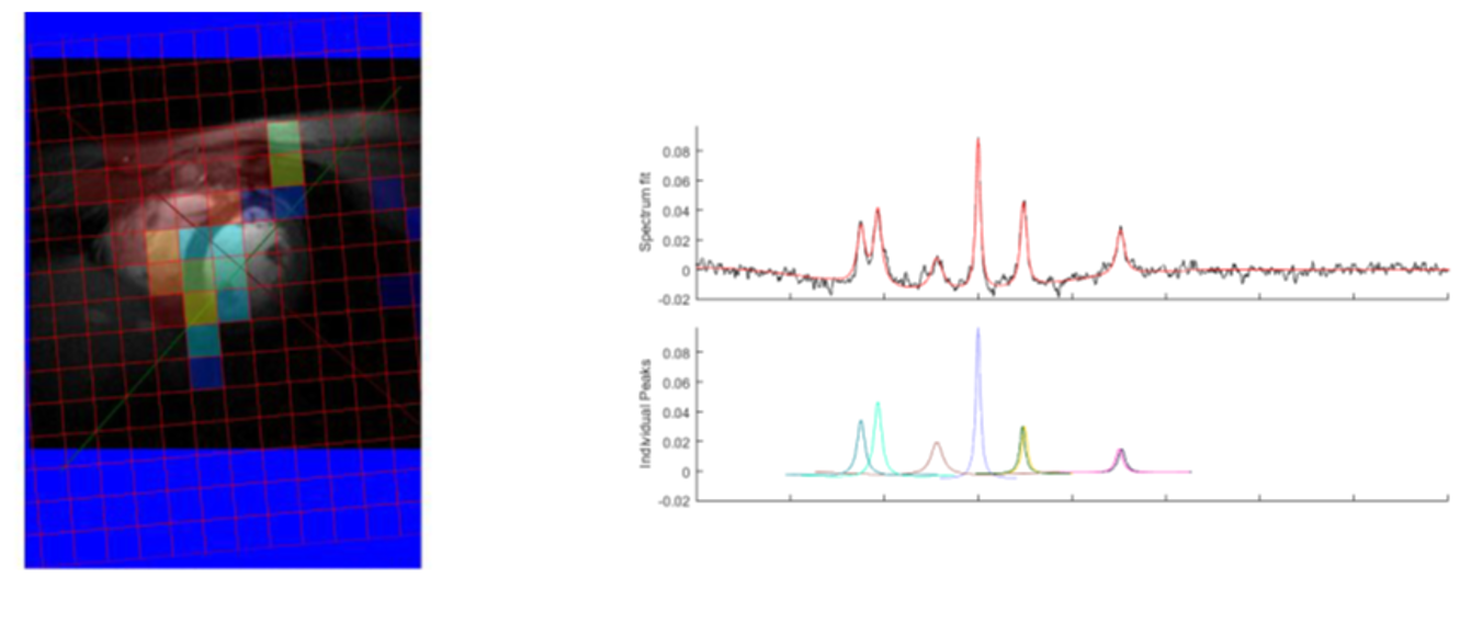
Figure 5: Results obtained in oxford with the array
16-channel coil. On the right, ATP concentration map overlaid on the heart
localizer for better visualization. On the left, an example of fitting using
AMARES algorithm (OXSA toolbox, MatLab). In the figure A there is a fitted
spectrum. In figure B individual peaks are depicted. Residuals are shown in
figure C and in the figure D the initial values for non-linear fit are
depicted.
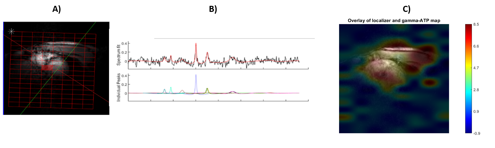
Figure 4: Results obtained in Cambridge with the dipole
array coil from Tesla DC. In A), the placement of the csi grid is shown. In B),
an example of fitting using AMARES algorithm (OXSA toolbox, MatLab) from the
highlighted pixel. In the figure, there is a fitted spectrum, individual peaks
are depicted. In C), The ATP concentration map is overlaid on the heart
localizer for better visualization.