Andrey N. Pravdivtsev1, Arne Brahms2, Jens Gröbner3, Rosa Rojas4, Eva Peschke1, Anna-Sophia Buschhoff4, Frowin Ellermann1, Rainer Herges2, Jan-Bernd Hövener1, and Peer Wulff4
1Section Biomedical Imaging, MOIN CC, Kiel University, Kiel, Germany, 2Otto Diels Institute for Organic Chemistry, Kiel University, Kiel, Germany, 3Fachhochschule Südwestfalen, Fachbereich Elektrotechnik und Informationstechnik, Lüdenscheid, Germany, 4Institute of Physiology, Kiel University, Kiel, Germany
1Section Biomedical Imaging, MOIN CC, Kiel University, Kiel, Germany, 2Otto Diels Institute for Organic Chemistry, Kiel University, Kiel, Germany, 3Fachhochschule Südwestfalen, Fachbereich Elektrotechnik und Informationstechnik, Lüdenscheid, Germany, 4Institute of Physiology, Kiel University, Kiel, Germany
We investigated the feasibility of 2H-MRS to assess the distribution of the anti-absence epilepsy drugs. The fast relaxation time and small magnetogyric ratio of deuterium allowed measuring of 2H-PRESS spectra of 4mm x 4mm x 4mm voxel with 9 mM of ethosuximide-D4,4' with SNR of 9 in 40 minutes.
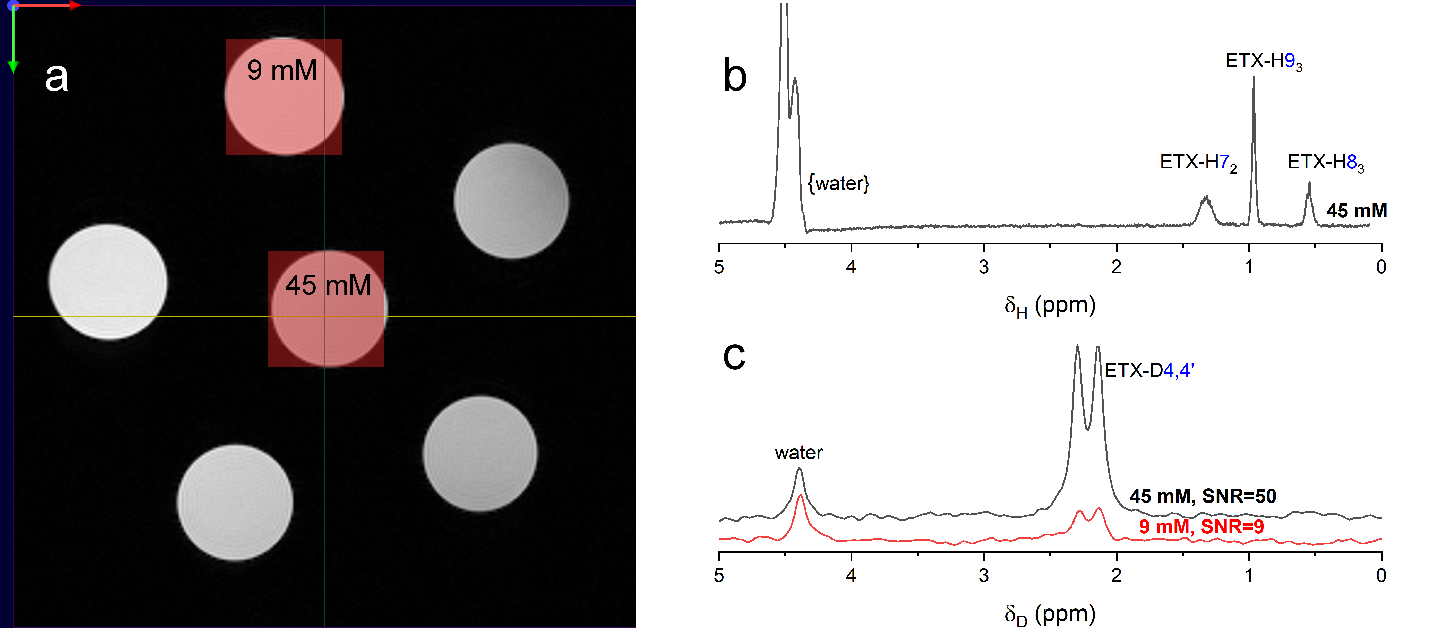
Figure 5. 1H
T2-TurboRARE image (a) 1H- (b) and 2H-PRESS spectra
(c). The phantom consists of six 5 mm NMR tubes with ETX-D4,4’
(0, 9, 18, 27, 36 and 45 mM) in aqueous 100 mM PBS buffer. Red boxes
represent the PRESS voxels: 4mm*4mm*4mm. 1H-PRESS parameters are:
TE=16.5ms, TR=2000ms, NS=1200, acquisition time AQ=467ms, experiment
time ET=40min, spectral bandwidth SW=4386 Hz. 2H-PRESS parameters are TE=14.2ms, TR=300ms, NS=8000, AQ=247ms, SW=8196Hz, ET=40min. Exponential
line broadening was 4 Hz. Spectra were zero-filled to achieved nominal spectral
resolution per point of 1 Hz.
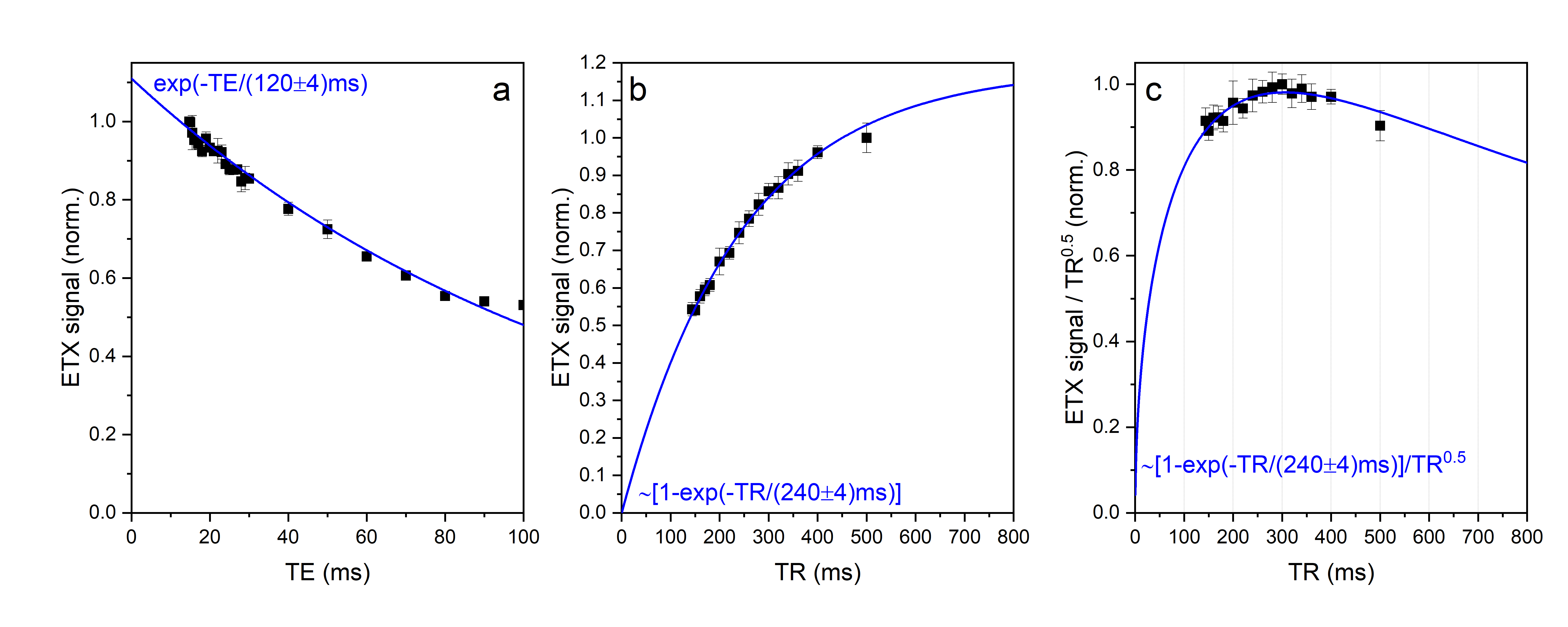
Figure 4. ETX-D4,4’ 2H-PRESS
signal as a function of TE (a, TR = 200 ms), TR (b, TE = 14.2 ms) and 2H-PRESS
signal divided by TR0.5 (c). The later reveals the most efficient for SNR value of TR with the fixed
scanning time. This value is predicted to be close to 1.25*T1 = 300 ms.
Lines are fitting of experimentally measured integrals of ETX-D4,4’, fitting
equations are given on the plots.