Stephanie J Blocker1, James Cook1, Yvonne M Mowery2, Jeffrey I Everitt3, Yi Qi1, Kathryn Hornburg1, Gary P Cofer1, Fernando Zapata1, Alex M Bassil2, Cristian T Badea1, David G Kirsch2, and G. Allan Johnson1
1Radiology, Duke University, Durham, NC, United States, 2Radiation Oncology, Duke University, Durham, NC, United States, 3Pathology, Duke University, Durham, NC, United States
1Radiology, Duke University, Durham, NC, United States, 2Radiation Oncology, Duke University, Durham, NC, United States, 3Pathology, Duke University, Durham, NC, United States
We have constructed a preclinical pipeline for registration of in vivo MRI, MR histology, and digitized pathology. Correlative analyses identified a selection of cytometric features in murine sarcomas which demonstrate linear trends with ex vivo and in vivo MR, including ADC and T2*.
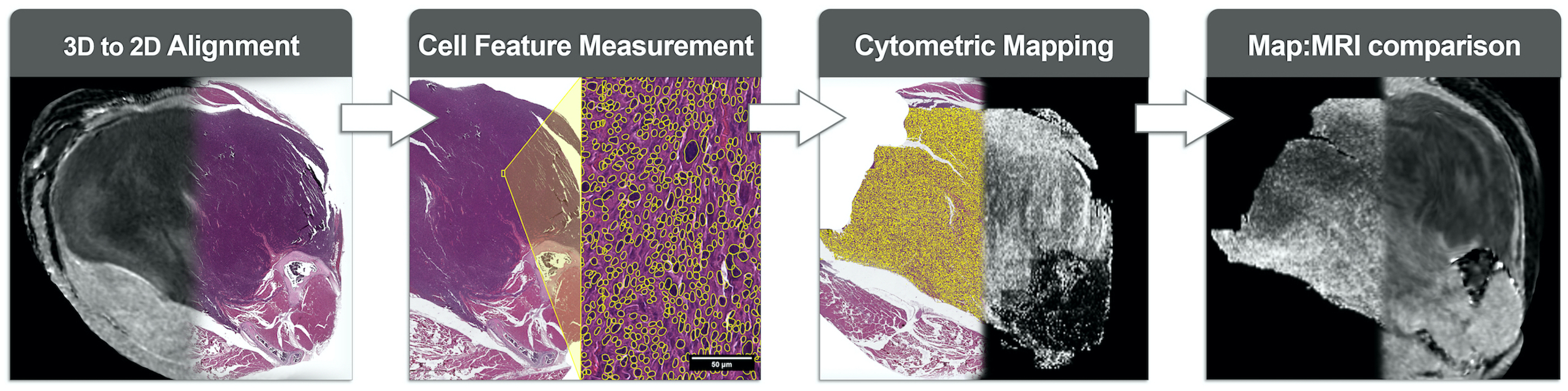
Figure 1: Schematic for co-registration of high-resolution 3D MRH with cytometric property maps derived from 2D H&E histology slides. Demonstration of the four phases for correlative MR studies: (1) Registration of MR to H&E slides; (2) Implementation of a multi-step algorithm for nuclear segmentation over entire histology slides; (3) Measurement of segmented nuclei and generation of quantitative cytometric feature maps; (4) Correlative studies of tumor MR signal and cytometric features.
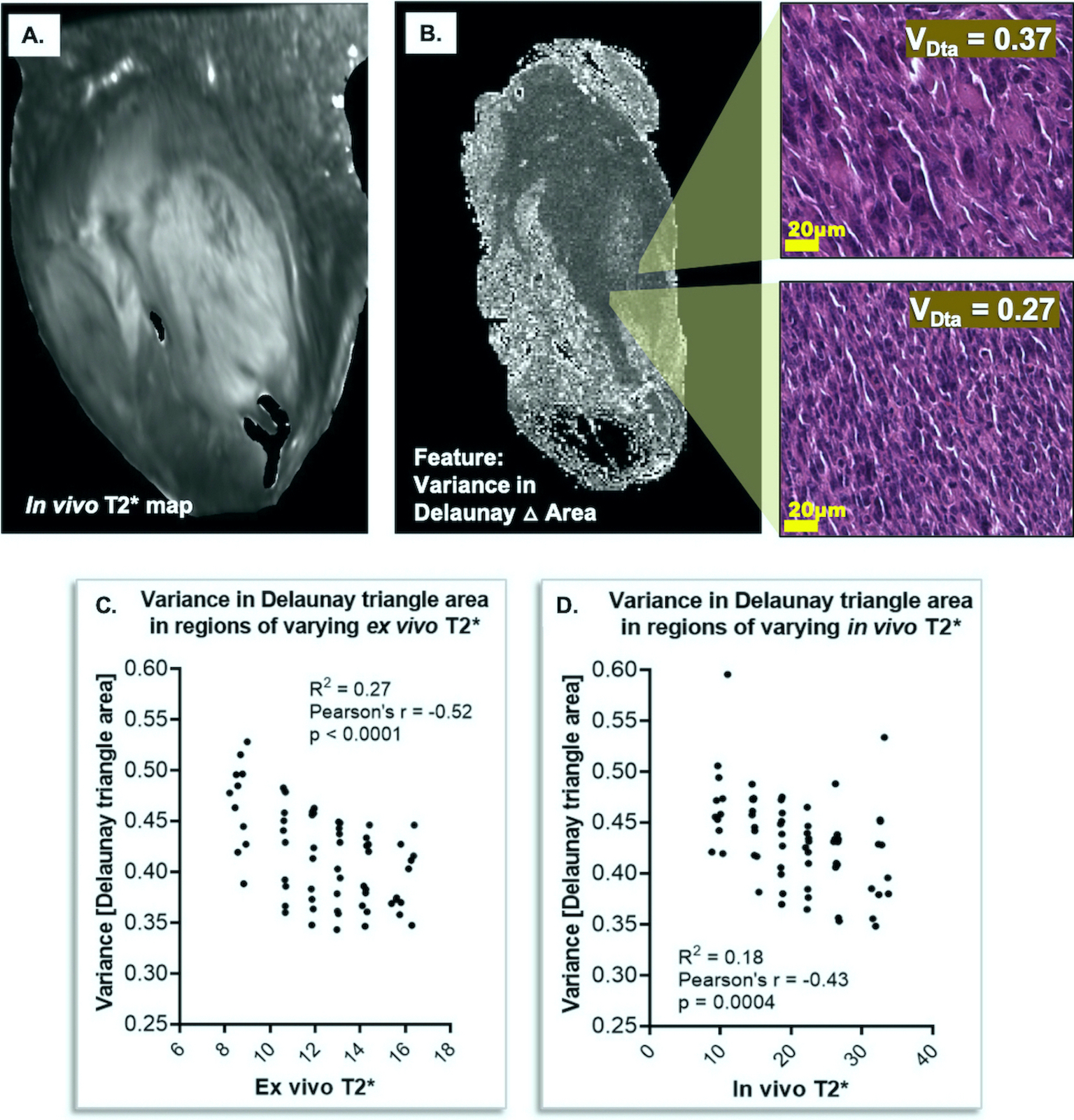
Figure 4: Relationships between MR and pathology features. Shown is an in vivo T2* sarcoma image (A). A map of variance in Delaunay triangle area (VDta) shows variable tumor cell organization (B). Correlation of VDta and T2* shows non-zero relationships in both ex vivo T2* (C; p < 0.0001, R2 = 0.27) and in vivo T2* (D; p = 0.0004, R2 = 0.18). R2 values reflect small pilot size (n=8).