Catherine Foss1, Desmond Jacob1, Flonné Wildes1, and Marie-France Penet1
1The Russell H. Morgan Department of Radiology and Radiological Science, The Johns Hopkins University School of Medicine, Baltimore, MD, United States
1The Russell H. Morgan Department of Radiology and Radiological Science, The Johns Hopkins University School of Medicine, Baltimore, MD, United States
Using PET-MRI in an orthotopic model of ovarian cancer, specific uptake of [124I]iodo-DPA-713 was observed in reactive macrophage, within and proximal to the primary tumor at early stages, and in ascitic fluid and lung metastases at later stages.
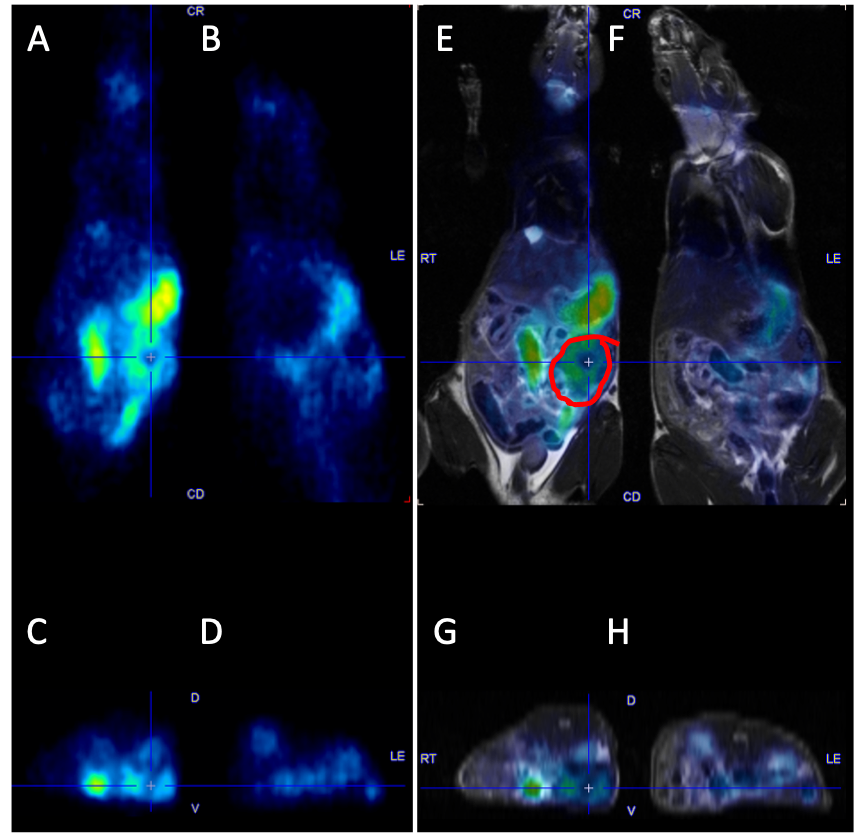
PET images of a tumor bearing mouse (A,C) and a sham mouse (B,D) with coronal (A,B) and corresponding axial slices (C,D). Corresponding fused PET-MR images (E-H).
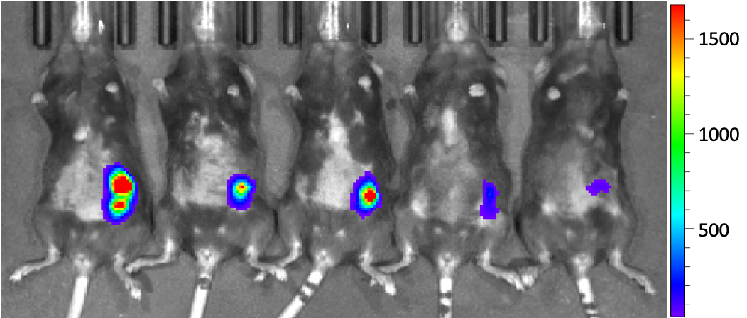
Bioluminescent images of 5 orthotopic ID8-Defb29-VEGF tumor bearing mice