Tianle Cao1,2, Sen Ma1, Nan Wang1, Sara Gharabaghi3, Yibin Xie1, Zhaoyang Fan1,4,5, Elliot Hogg6, E. Mark Haacke 3,7,8, Michele Tagliati6, Anthony G. Christodoulou1,2, and Debiao Li1,2
1Biomedical Imaging Research Institute, Cedars Sinai Medical Center, Los Angeles, CA, United States, 2Department of Bioengineering, University of California, Los Angeles, Los Angeles, CA, United States, 3Magnetic Resonance Innovations, Inc., Bingham Farms, MI, United States, 4Department of Radiation Oncology, Keck School of Medicine, University of Southern California, Los Angeles, CA, United States, 5Department of Radiology, University of Southern California, Los Angeles, CA, United States, 6Department of Neurology, Cedars Sinai Medical Center, Los Angeles, CA, United States, 7Department of Radiology, Wayne State University School of Medicine, Detroit, MI, United States, 8The MRI Institute for Biomedical Research, Bingham Farms, MI, United States
1Biomedical Imaging Research Institute, Cedars Sinai Medical Center, Los Angeles, CA, United States, 2Department of Bioengineering, University of California, Los Angeles, Los Angeles, CA, United States, 3Magnetic Resonance Innovations, Inc., Bingham Farms, MI, United States, 4Department of Radiation Oncology, Keck School of Medicine, University of Southern California, Los Angeles, CA, United States, 5Department of Radiology, University of Southern California, Los Angeles, CA, United States, 6Department of Neurology, Cedars Sinai Medical Center, Los Angeles, CA, United States, 7Department of Radiology, Wayne State University School of Medicine, Detroit, MI, United States, 8The MRI Institute for Biomedical Research, Bingham Farms, MI, United States
A new approach for simultaneous quantitative mapping of T1, T2, T2*, and susceptibility was developed. Results of both visual comparison and statistical analysis showed that our proposed method agreed well with reference methods while being more time efficient.
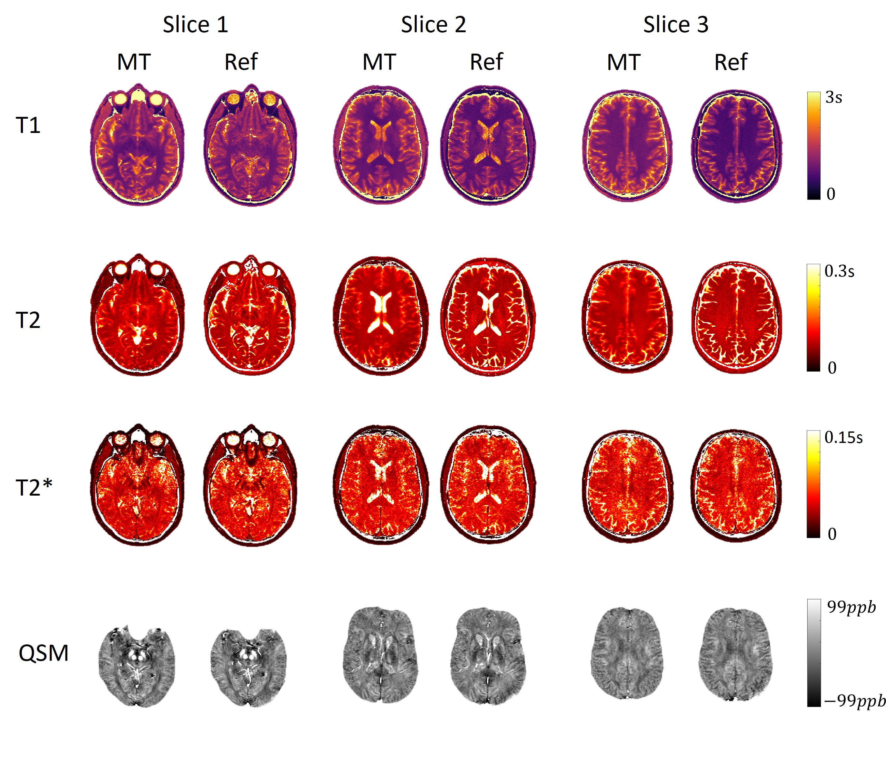
Figure 2. Representative T1/T2/T2*/QSM mapping at
3 slice locations using MR Multitasking and the corresponding reference
protocols on a healthy volunteer.
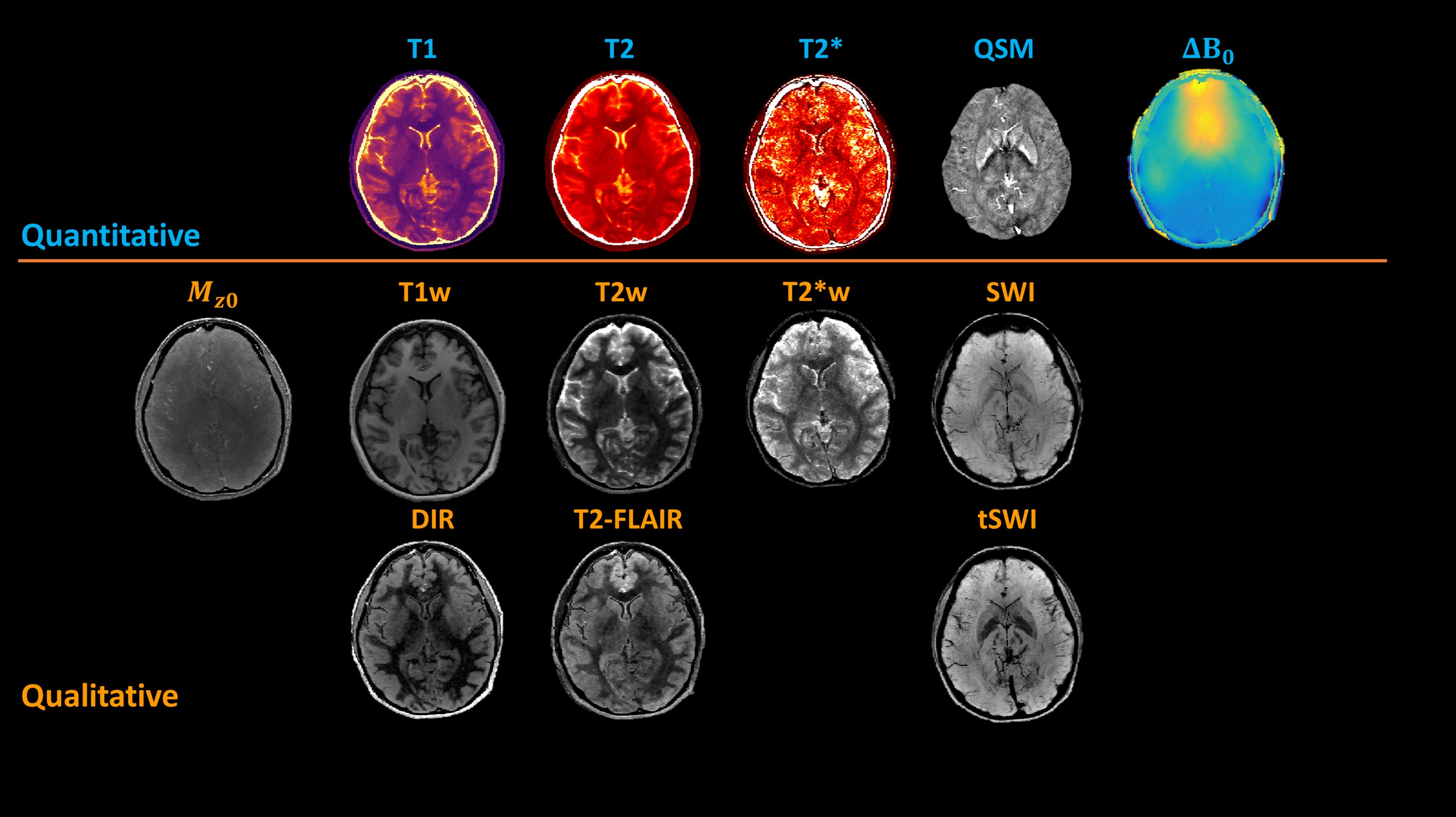
Figure 4. Results from a
23-year-old female volunteer including qualitative images and quantitative
data. The first row shows quantitative maps and the second and third rows show the
synthesized weighted images. SWI and tSWI were minimum intensity projection (mIP) results with an effective
slab thickness of 16 mm. χ1=0, χ2=450ppb was adopted for tSWI23.