Giorgia Milotta1, Gastao Cruz2, Radhouene Neji2, Claudia Prieto2, and Rene Botnar2
1University College London, London, United Kingdom, 2King's College London, London, United Kingdom
1University College London, London, United Kingdom, 2King's College London, London, United Kingdom
The proposed approach
permits the acquisition of 3D free-breathing T1, T2 and fat fraction maps in a scan
time of less than 6 minutes. Increased sharpness and reduced spatial
variability was observed with non-rigid motion correction.
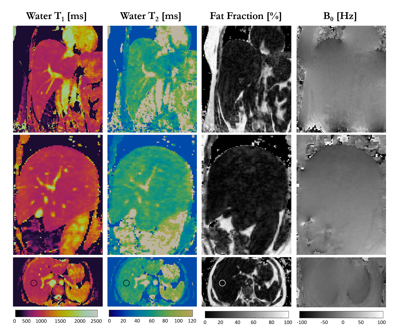
Figure 3
– Co-registered T1 and T2 maps, fat fraction and B0 map for one healthy subject
acquired with 3-point Dixon GRE read out. Coronal, sagittal and transversal
views are shown. T1=704±30ms, T2 = 58±3ms and fat fraction = 3.3±1.7% were
measured within a ROI in the liver. Acquisition parameters included FA=8deg,
isotropic resolution of 2mm3, FOV=320x320x168mm3, coronal
orientation, 14 echoes for iNAV acquisition, acquisition window of 200ms,
bandwidth=801Hz/pixel, T2prep=50ms, TI=120ms and total scan time of ~6min.
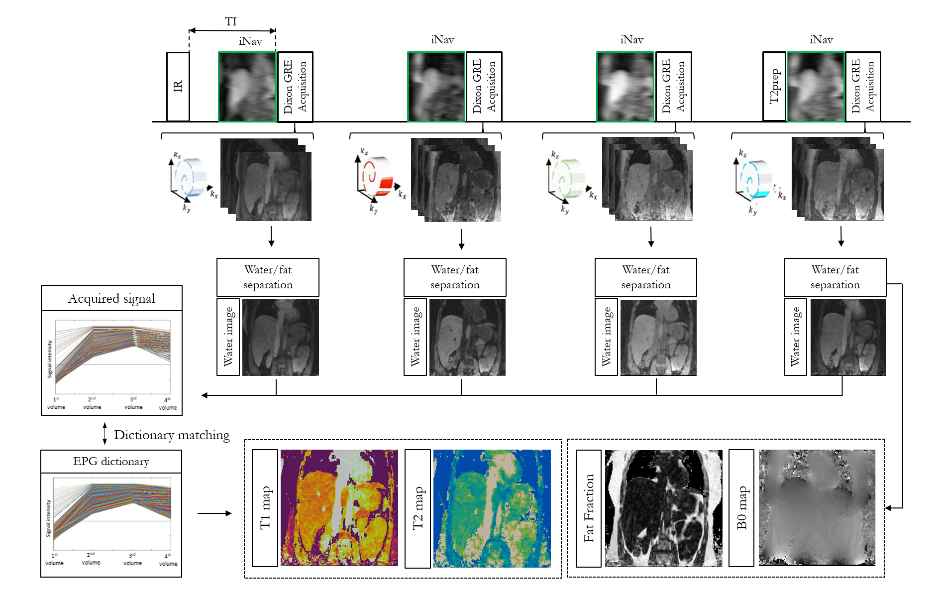
Figure 1 – Four
interleaved volumes are acquired with variable density Cartesian trajectory, three-point
Dixon GRE readout. 2D-iNAVs are acquired to correct for translational motion
and respiratory binning. Water images are generated with Dixon water/fat separation
and used to obtain the signal evolution of each voxel. T1 and T2
maps are obtained by matching the acquired signal evolution to the EPG
simulated dictionary. Fat fraction and M0 map are obtained from water/fat
separation algorithm.