Rakshit Dadarwal1,2 and Susann Boretius1,2
1Functional Imaging Laboratory, German Primate Center, Göttingen, Germany, 2Georg August Universität Göttingen, Göttingen, Germany
1Functional Imaging Laboratory, German Primate Center, Göttingen, Germany, 2Georg August Universität Göttingen, Göttingen, Germany
We provide a high-resolution multi-contrast MRI template space for the cynomolgus macaque brain in the stereotaxic space. Template space also offers cortical, subcortical, and white matter structural parcellation.
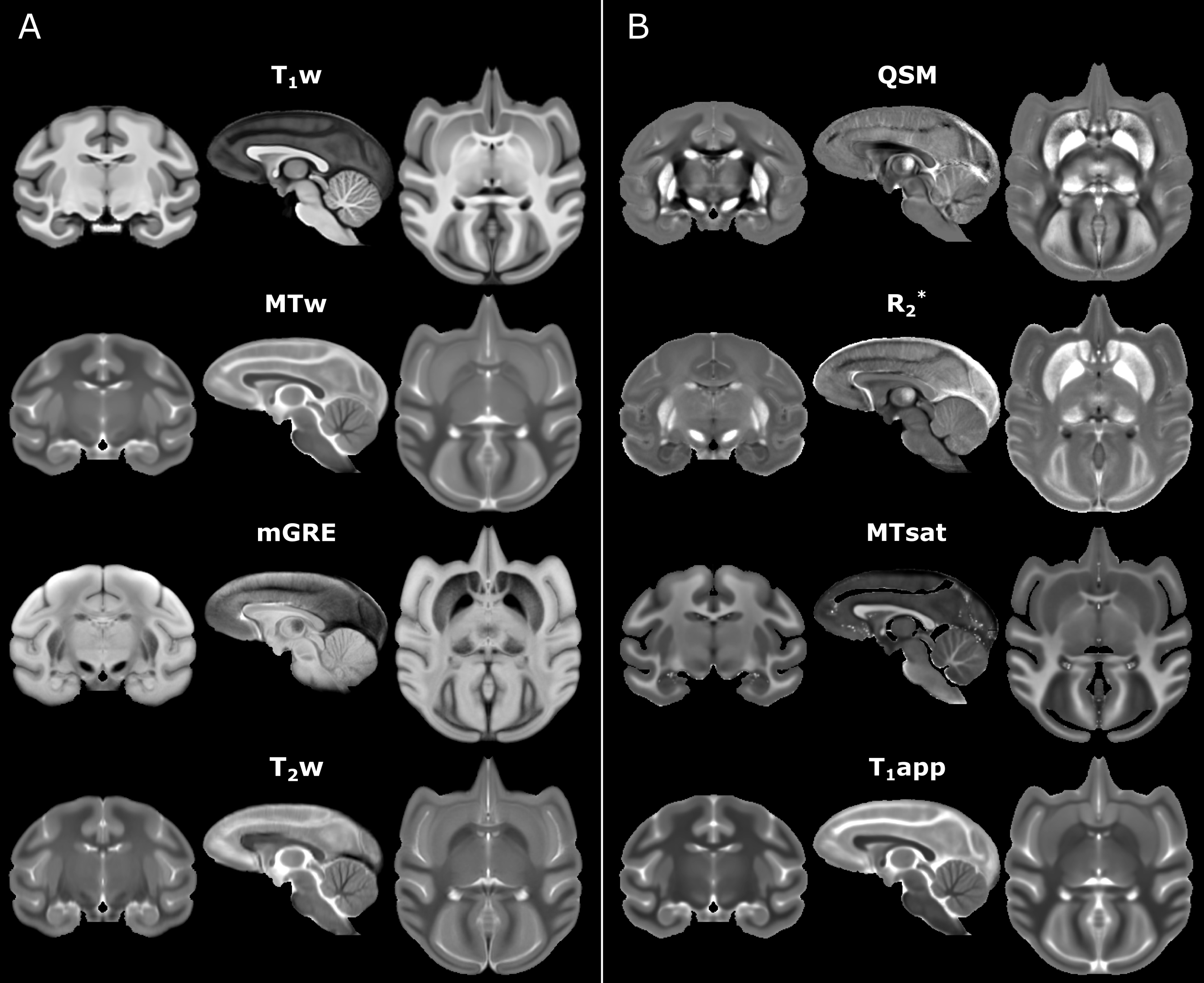
Figure 1.
The symmetric cynomolgus macaque brain templates in coronal, sagittal, and axial view. A) Templates of the originally acquired weighted data sets (from top to bottom) include T1-weighted, MT-weighted, ME-GRE mean across echo times (mGRE), and T2-weighted template. B) Parametric map templates (from top to bottom) incorporate QSM, R2*, and MT saturation and apparent T1 maps.
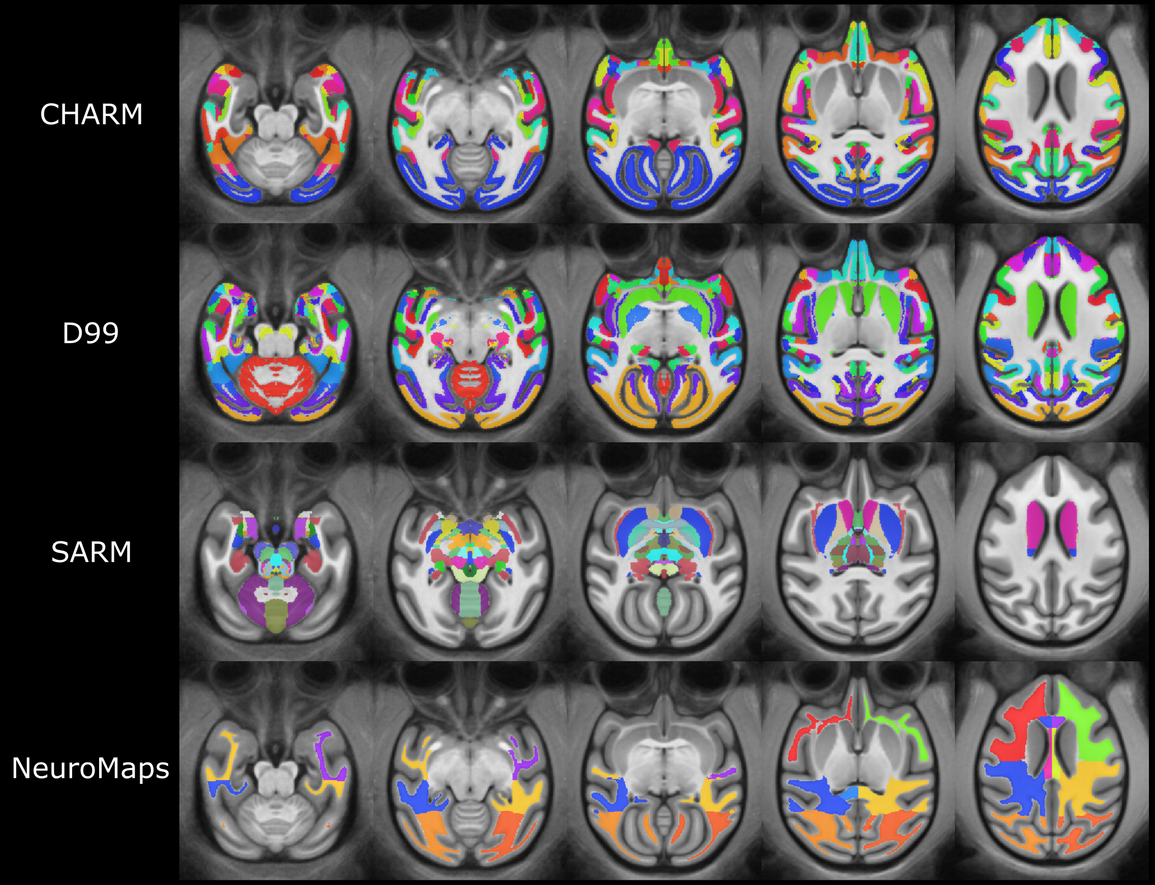
Figure 2. Cynomolgus macaque brain structural parcellation derived from the openly available rhesus macaque brain templates. Emanated ROI labels are from the Cortical Hierarchy Atlas of the Rhesus Macaque (CHARM), D99, Subcortical Atlas of the Rhesus Macaque (SARM), and NeuroMaps atlas.