Jing Liu1, Angela Jakary1, Duan Xu1, and Janine Lupo1
1Radiology and Biomedical Imaging, University of California San Francisco, San Francisco, CA, United States
1Radiology and Biomedical Imaging, University of California San Francisco, San Francisco, CA, United States
Optimization of the imaging parameters and motion compensation for multi-contrast whole brain MRI in a single 3D acquisition resulted in imporoved image quality and will benefit for better accuracy of the resulting brain tissue segmentation and lesion detection.
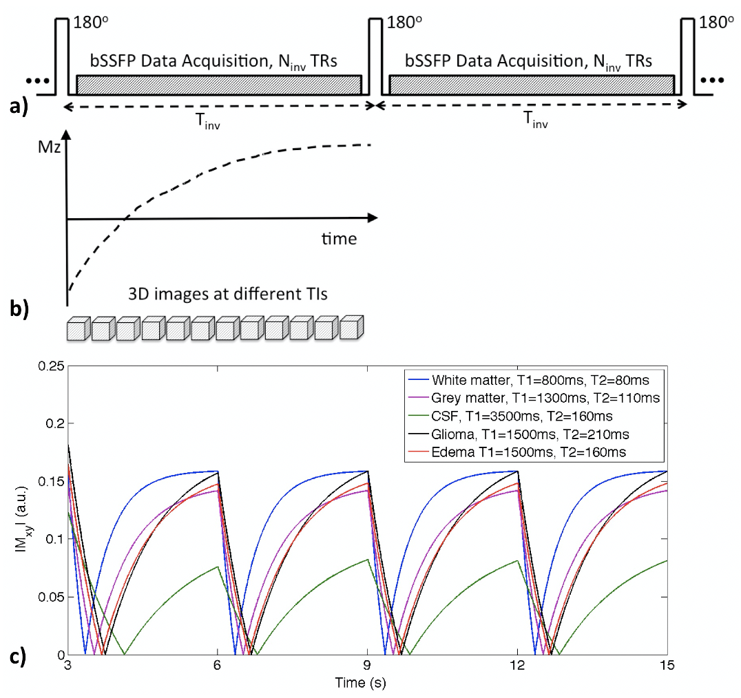
Figure 1. a) IIR-bSSFP acquisition. A nonselective inversion pulse is applied followed by a fixed number of segmented bSSFP acquisitions (interval of Tinv). b) With a continuous data acquisition, a series of 3D images are reconstructed at different TIs. c) Simulated signal evolutions of different brain tissues with assumed T1/T2 values and scan parameters (Tinv=3s, flip angle=30 degrees, TR=4ms).
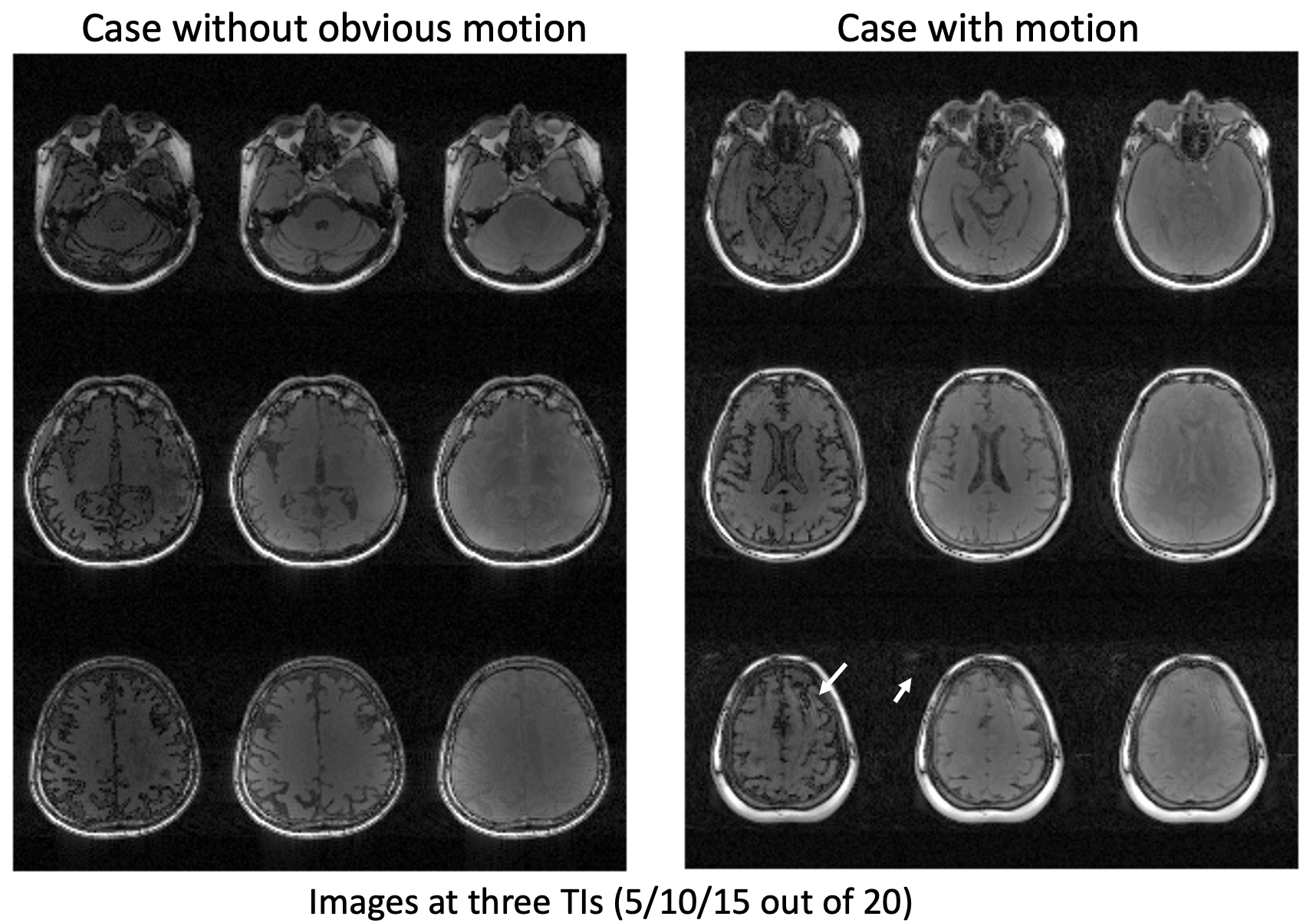
Figure 3. Whole brain multi-contrast MRI acquired in a single scan of 6 minutes. The imaging sequence has inherent motion robustness to small amounts of motion, but any excess motion would cause image quality degradation without motion compensation (right block, arrows highlighting motion artifacts).