Rakshit Dadarwal1,2 and Susann Boretius1,2
1Functional Imaging Laboratory, German Primate Center, Göttingen, Germany, 2Georg August Universität Göttingen, Göttingen, Germany
1Functional Imaging Laboratory, German Primate Center, Göttingen, Germany, 2Georg August Universität Göttingen, Göttingen, Germany
We have proposed a method to combine T1w and QSM contrasts that significantly improves the quality of subcortical nuclei classification while retaining the excellent white matter delineation of T1w.
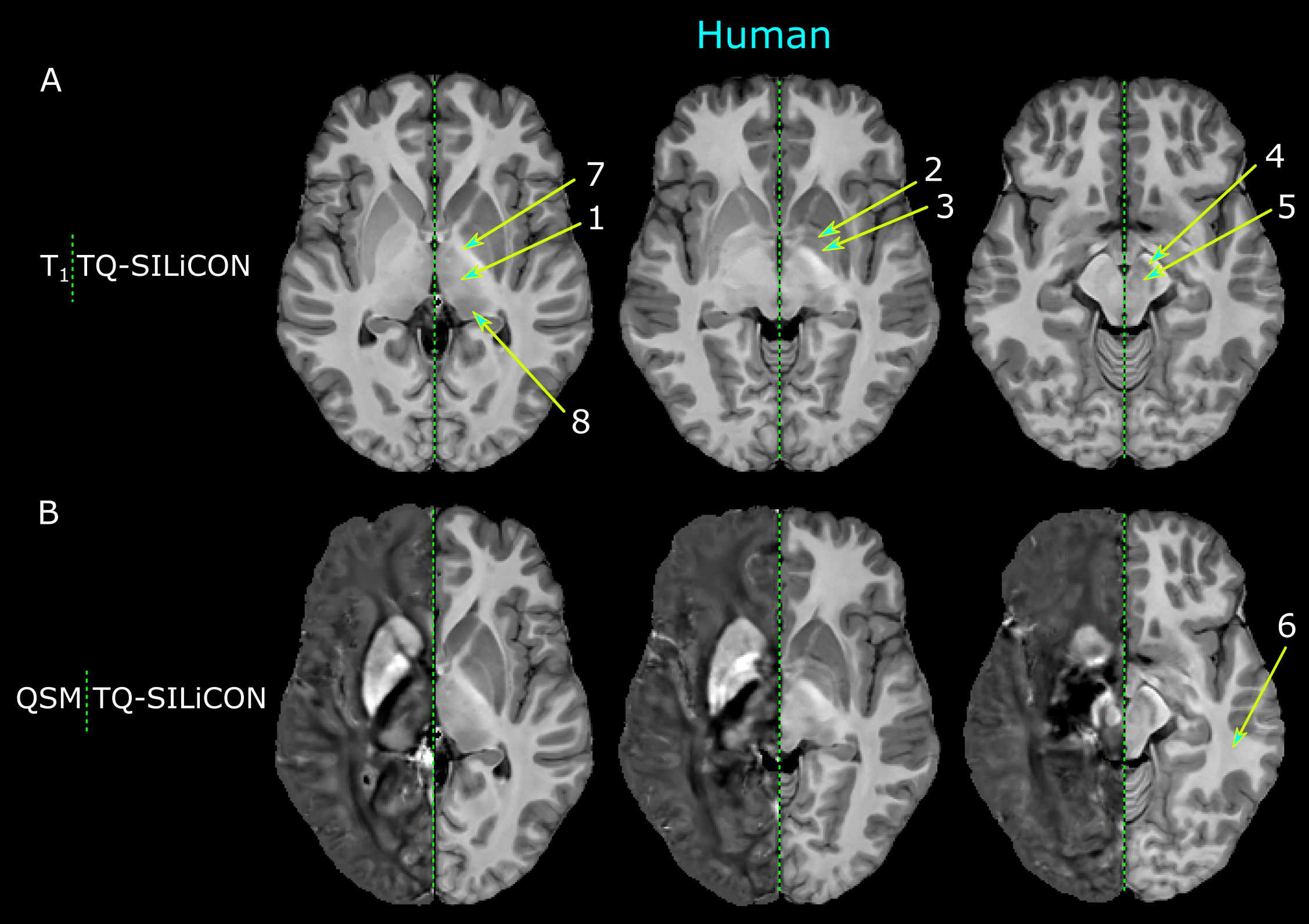
Figure 1. The juxtaposition of a human subject’s T1–QSM fusion image (TQ-SILiCON map) in comparison with the respective T1 weighted image (A, left) and QSM (B, left). TQ-SILiCON maps were generated with the weights of 0.318 and -0.790 for T1w and QSM respectively. 1 – thalamus, 2 – external globus pallidus, 3 – internal globus pallidus, 4 –substantia nigra, 5 – red nucleus, 6 – white matter, 7 – internal capsule, and 8 – pulvinar nucleus.
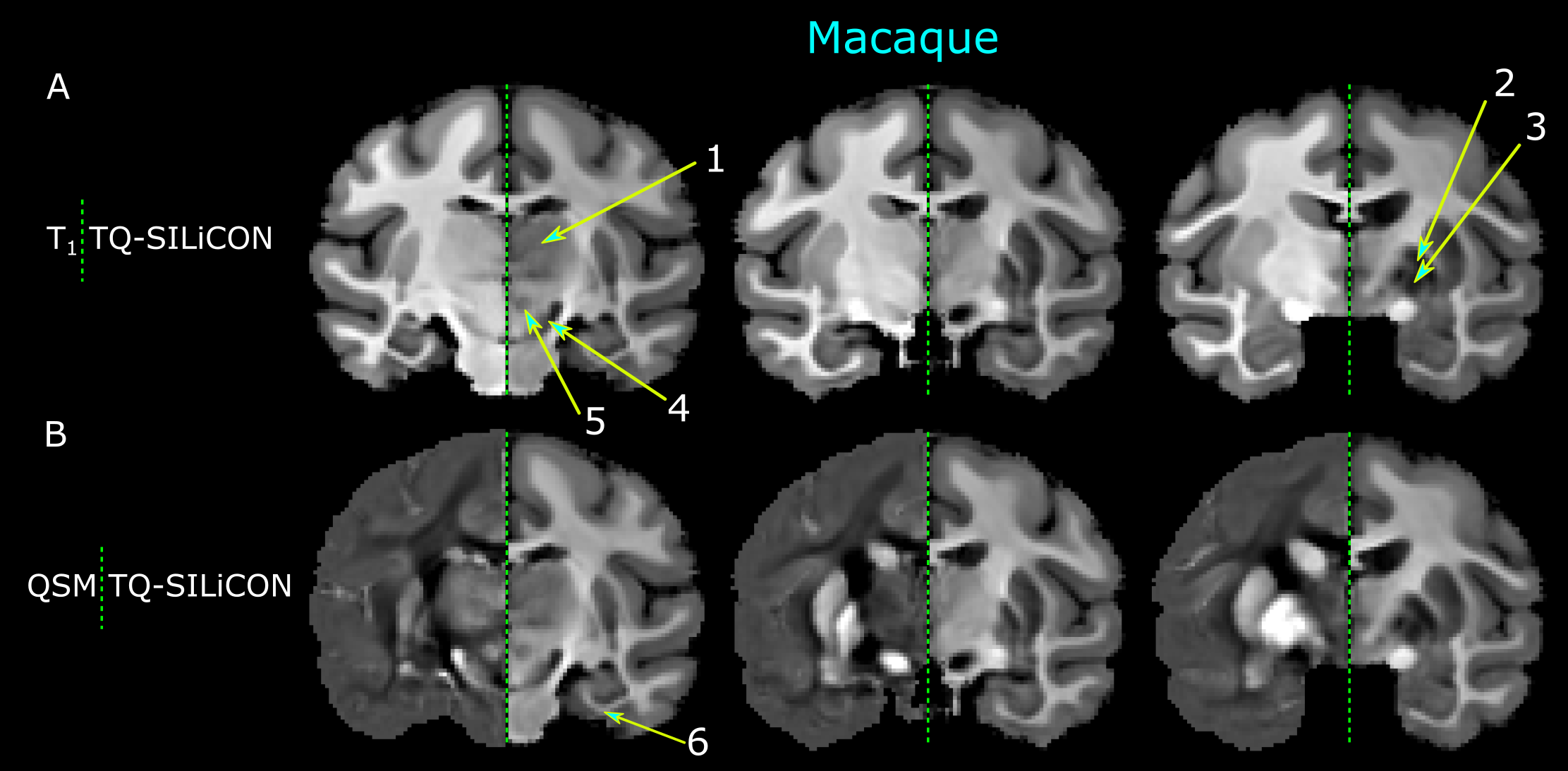
Figure 2. Illustration of a macaque’s T1-QSM fusion image (TQ-SILiCON map) in comparison with the respective T1 weighted image (A, left) and QSM (B, left). The emphasized areas on the TQ-SILiCON map are parts of the subcortical gray matter nuclei: 1 – thalamus, 2 – external globus pallidus, 3 – internal globus pallidus, 4 – substantia nigra, 5 – red nucleus, and 6 – white matter.