Weijian Huang1, Yulon Qi2, Qiang He3, Ting Ma4, Xin Liu1, Guanxun Cheng2, Hairong Zheng1, and Shanshan Wang1
1Paul C Lauterbur Research Center, Shenzhen Institutes of Advanced Technology Chinese Academy of Sciences, Shenzhen, China, 2Radiology department, Peking University Shenzhen Hospital, shenzhen, China, 3United Imaging Research Institute of Innovative Medical Equipment, Shenzhen, China, 4Pengcheng Laboratory, Shenzhen, China
1Paul C Lauterbur Research Center, Shenzhen Institutes of Advanced Technology Chinese Academy of Sciences, Shenzhen, China, 2Radiology department, Peking University Shenzhen Hospital, shenzhen, China, 3United Imaging Research Institute of Innovative Medical Equipment, Shenzhen, China, 4Pengcheng Laboratory, Shenzhen, China
Identifying stroke lesions is crucial in clinic. The misalignment between multi-contrast images is the main obstacle in this process. This paper proposes a new registration framework to for accurate and quick multi-contrast image registration, with promising performances achieved.

Fig. 3. Qualitative results of different methods. The blue and red edge lines indicate the stroke lesion regions annotated by radiologists based on DWI and FLAIR, and the green lines indicate the predictions of different methods.
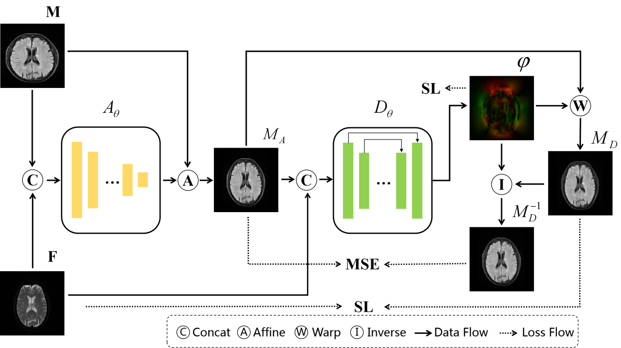
Fig.2. Flow chart of the proposed method. M represents the moving image, F represents the fixed image, and M-1D represents the prediction of the inverse transformation. We impose MSE constraints on M-1D and MA to ensure topological deformation.