Thomas Campbell Arnold1, Samantha By2, Hadrien Dyvorne2, Rafael O'Halloran2, Farzana Sayani3, Lisa M. Desiderio4, Brian Litt1,5, and Joel M. Stein4
1Bioengineering, University of Pennsylvania, Philadelphia, PA, United States, 2Hyperfine Research, Guilford, CT, United States, 3Medicine, Perelman School of Medicine, Philadelphia, PA, United States, 4Radiology, Perelman School of Medicine, Philadelphia, PA, United States, 5Neurology, Perelman School of Medicine, Philadelphia, PA, United States
1Bioengineering, University of Pennsylvania, Philadelphia, PA, United States, 2Hyperfine Research, Guilford, CT, United States, 3Medicine, Perelman School of Medicine, Philadelphia, PA, United States, 4Radiology, Perelman School of Medicine, Philadelphia, PA, United States, 5Neurology, Perelman School of Medicine, Philadelphia, PA, United States
Ferumoxytol
is a promising contrast agent in low-field MRI. We characterize ferumoxytol
T1/T2 relaxation properties and collect in-vivo imaging of iron-deficiency
anemia patients. We observed contrast enhancement on T1/T2/FLAIR imaging and venous/arterial
contrast in an angio sequence.
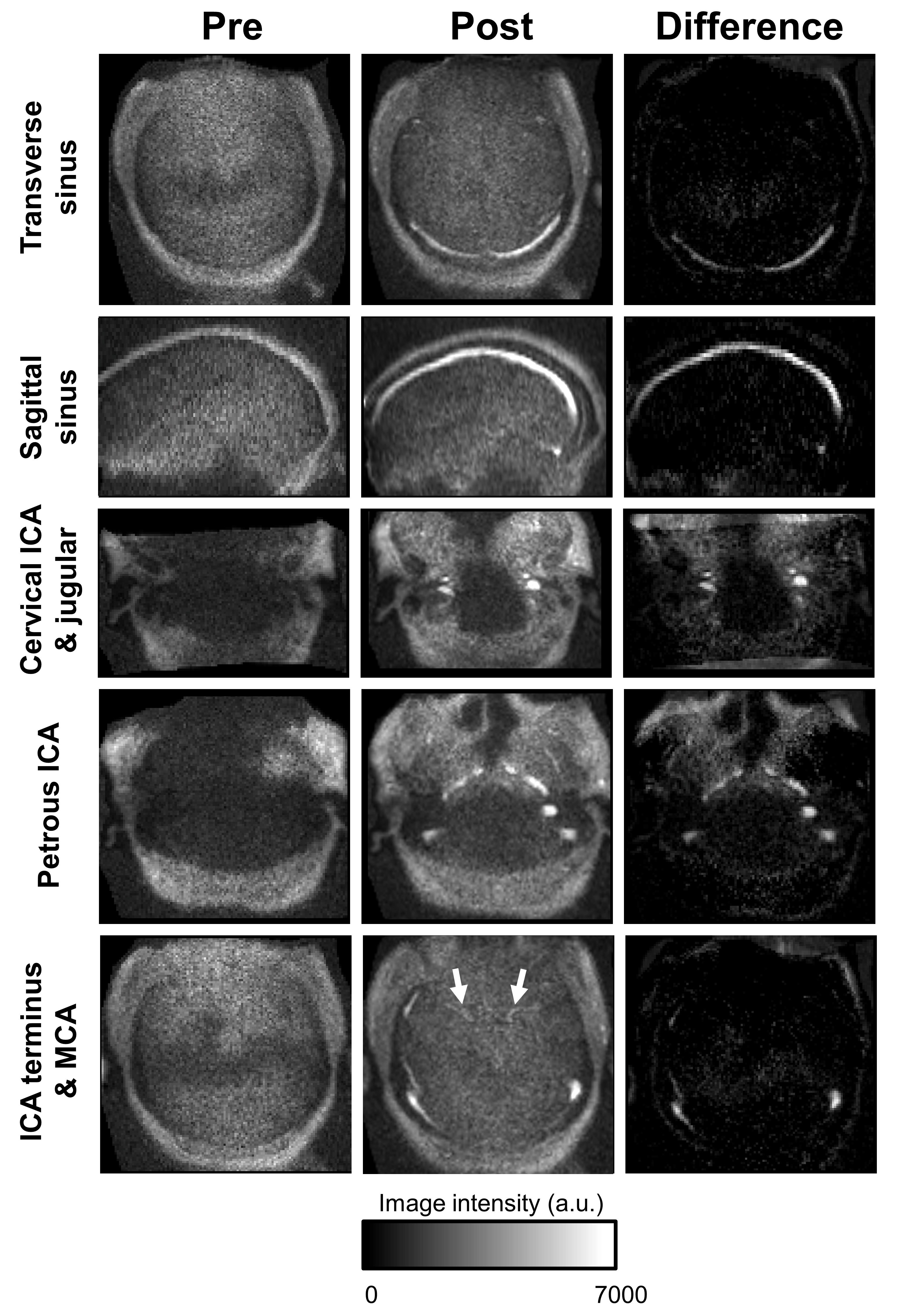
T1-weighted angiographic imaging with ferumoxytol at 64mT showing
enhancement of dural venous sinuses and jugular veins, distal cervical and
intracranial internal carotid arteries (ICA) as well as middle cerebral
arteries (MCA, arrows).
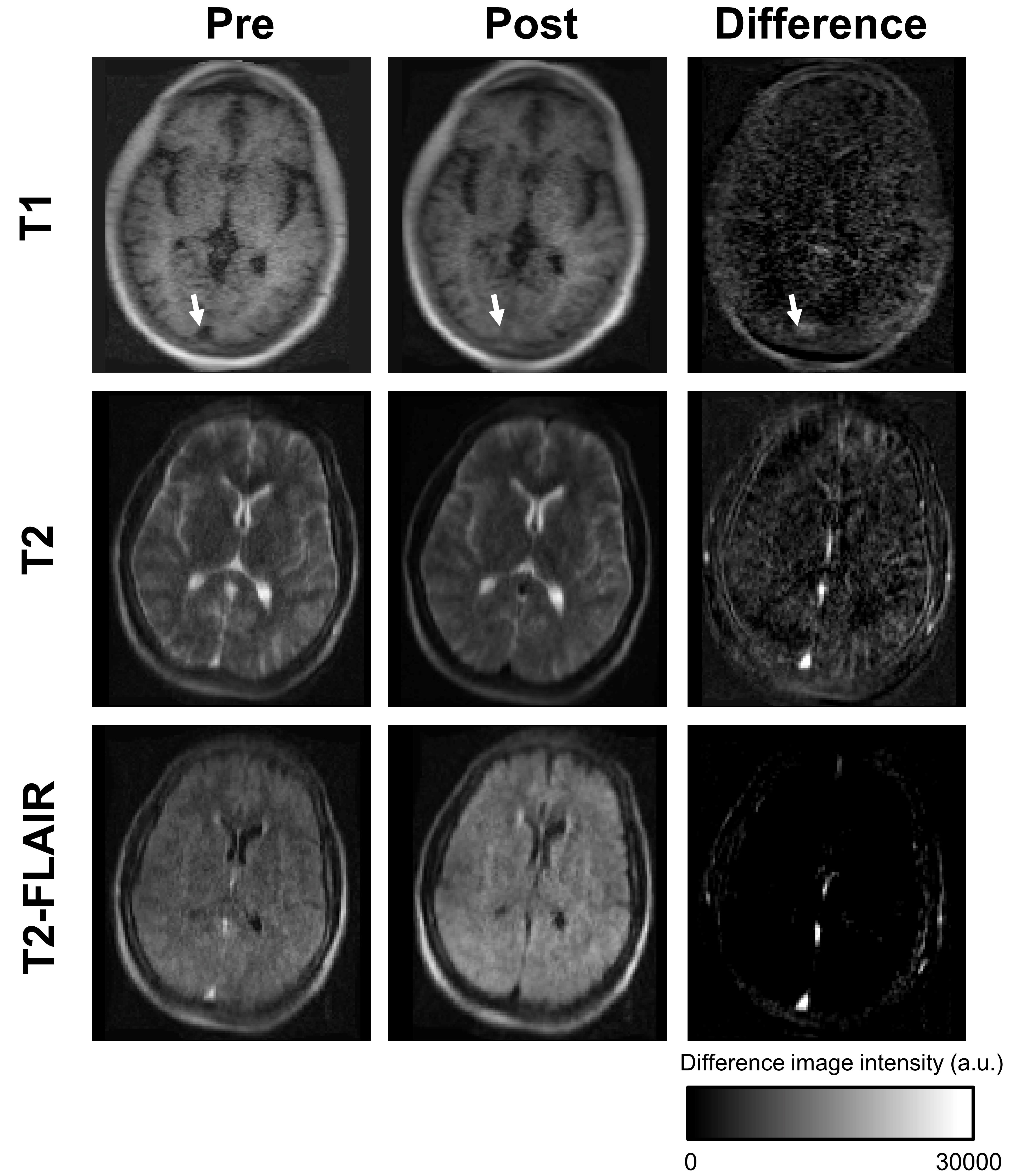
Contrast
enhancement with ferumoxytol on standard sequences optimized for brain tissue
contrasts at 64 mT. On T1 images, ferumoxutol modestly increases signal in the superior
sagittal sinus (arrows) and other venous structures. On T2 and T2-FLAIR images
ferumoxytol markedly decreases signal of intrinsically hyperintense veins, shown
as enhancement on inverse difference images (i.e. pre-minus-post).