Fábio Seiji Otsuka1, Carlos Salmon1, Otaciro Nasimento2, and Maria Otaduy3
1Physics Department, University of São Paulo, Ribeirão Preto, Brazil, 2Physics Institute, University of São Paulo, São Carlos, Brazil, 3Medical School, University of São Paulo, São Paulo, Brazil
1Physics Department, University of São Paulo, Ribeirão Preto, Brazil, 2Physics Institute, University of São Paulo, São Carlos, Brazil, 3Medical School, University of São Paulo, São Paulo, Brazil
Four different peaks were observed on all brain regions. The rhombic iron, copper and organic radical peaks presented a Curie behavior while the broad peak at g = 2.0 presented an antiferromagnetic behavior. Finally, the Locus Coeruleus showed an additional peak.
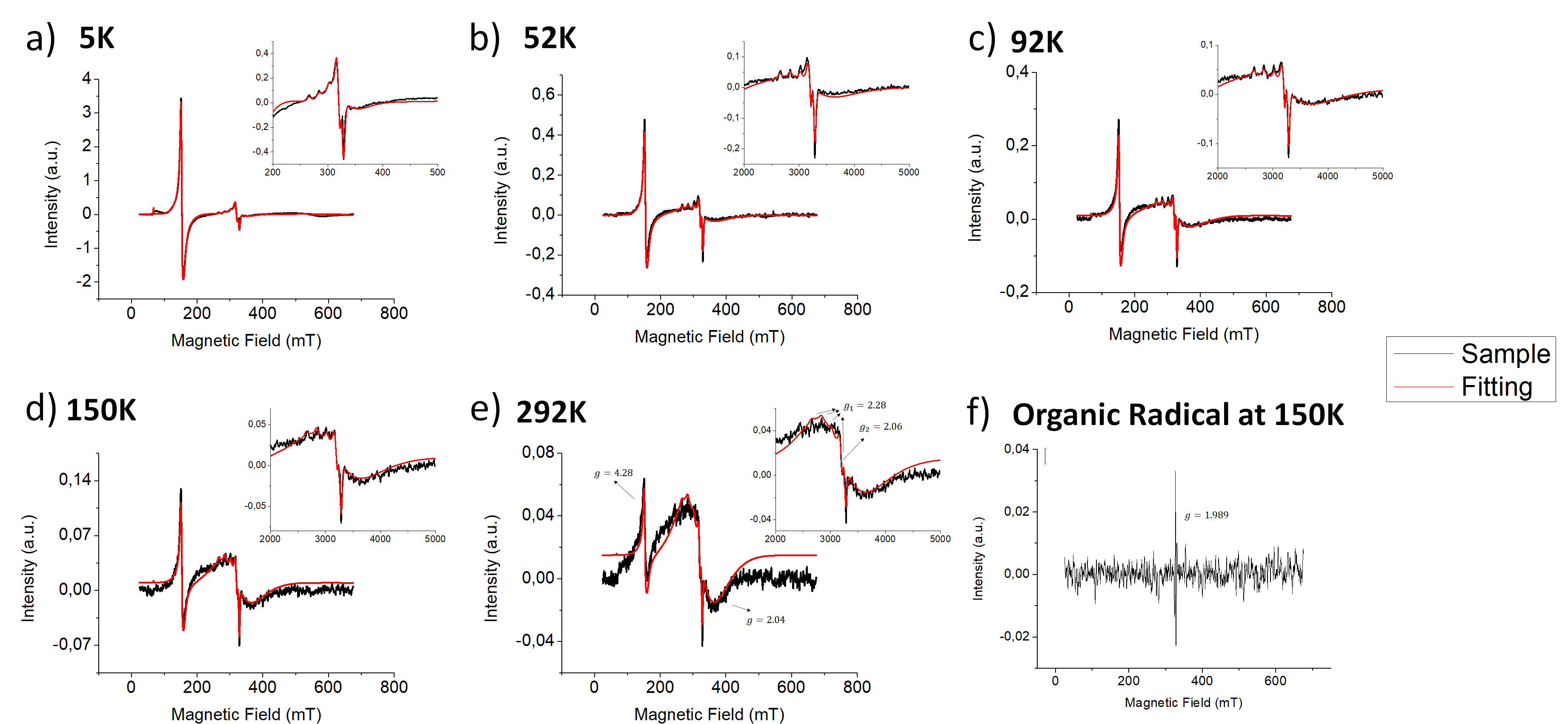
Figure 1 –
(a-e) EPR spectra and the fitting of GP sample at different temperatures.
Inserts represent the spectrum region around g = 2.0. f) The organic radical
peak after subtraction of other fitted peaks.
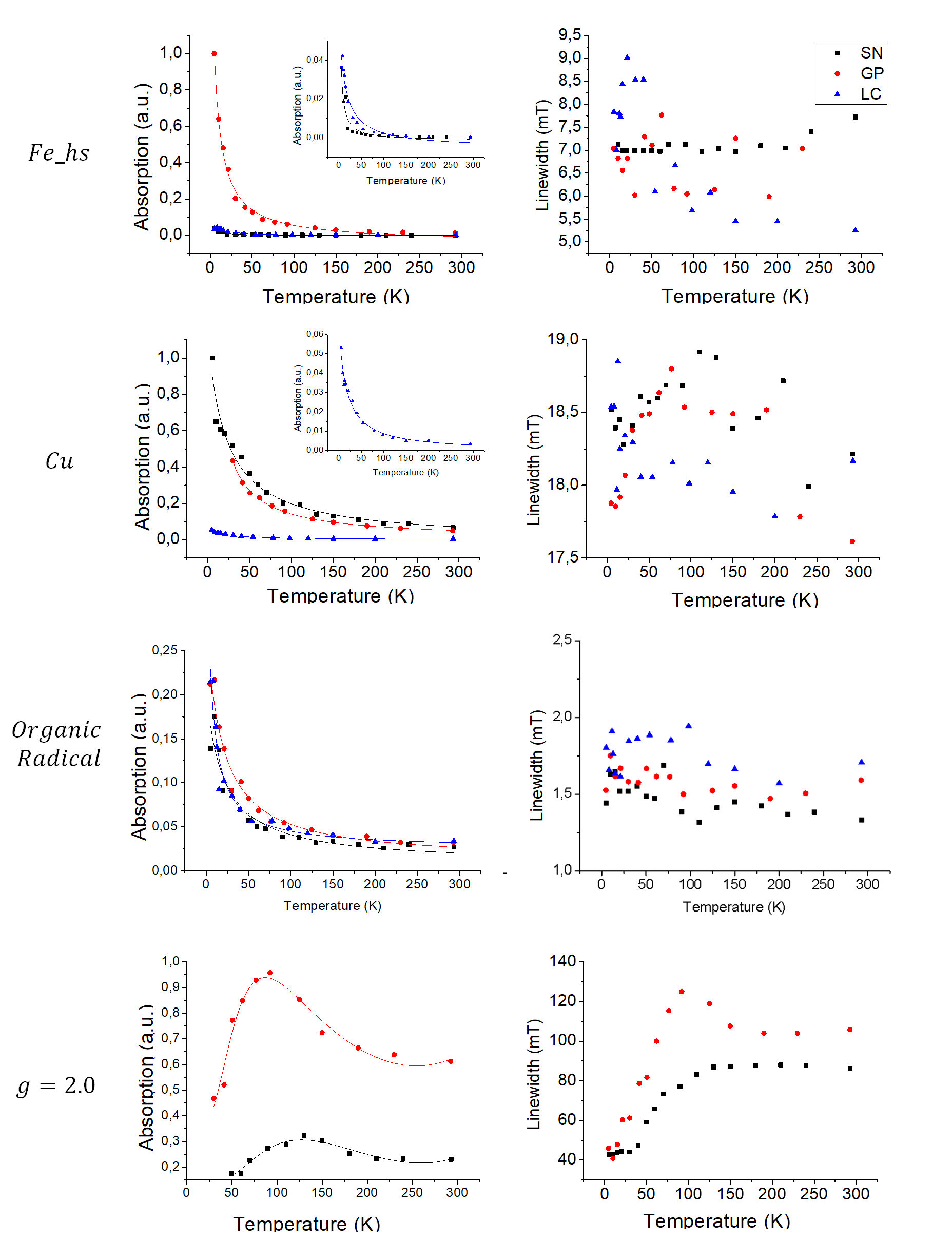
Figure 2 –
Second integral and linewidth as a function of temperature for each peak
(Fe_hs, Cu, broad peak at g=2.0 and the organic radical) and each sample (SN,
GP, LC). For the Fe_hs, Cu and organic radical peaks fitting was performed
using Curie-Weiss Law. For the broad peak Vav-Vleck paramagnetism for S=5/2 and
S=1/2 with Curie-Weiss law was used for fitting.