Caroline Le Ster1, Franck Mauconduit1, Christian Mirkes2, Michel Bottlaender3,4, Fawzi Boumezbeur1, Boucif Djemai1, Alexandre Vignaud1, and Nicolas Boulant1
1Paris-Saclay University, CEA, CNRS, BAOBAB, Neurospin, Gif-sur-Yvette, France, 2Skope MRT, Zurich, Switzerland, 3Paris-Saclay University, CEA, CNRS, INSERM, BioMaps, Service hospitalier Joliot, Orsay, France, 4UNIACT, Neurospin, CEA, Gif-sur-Yvette, France
1Paris-Saclay University, CEA, CNRS, BAOBAB, Neurospin, Gif-sur-Yvette, France, 2Skope MRT, Zurich, Switzerland, 3Paris-Saclay University, CEA, CNRS, INSERM, BioMaps, Service hospitalier Joliot, Orsay, France, 4UNIACT, Neurospin, CEA, Gif-sur-Yvette, France
Inclusion of field fluctuations in MR thermometry experiments performed at 7T with concurrent radiofrequency heating on an agar-gel phantom and on an anaesthetized macaque showed beneficial for the measurement of small temperature rises as encountered in standard brain exams.
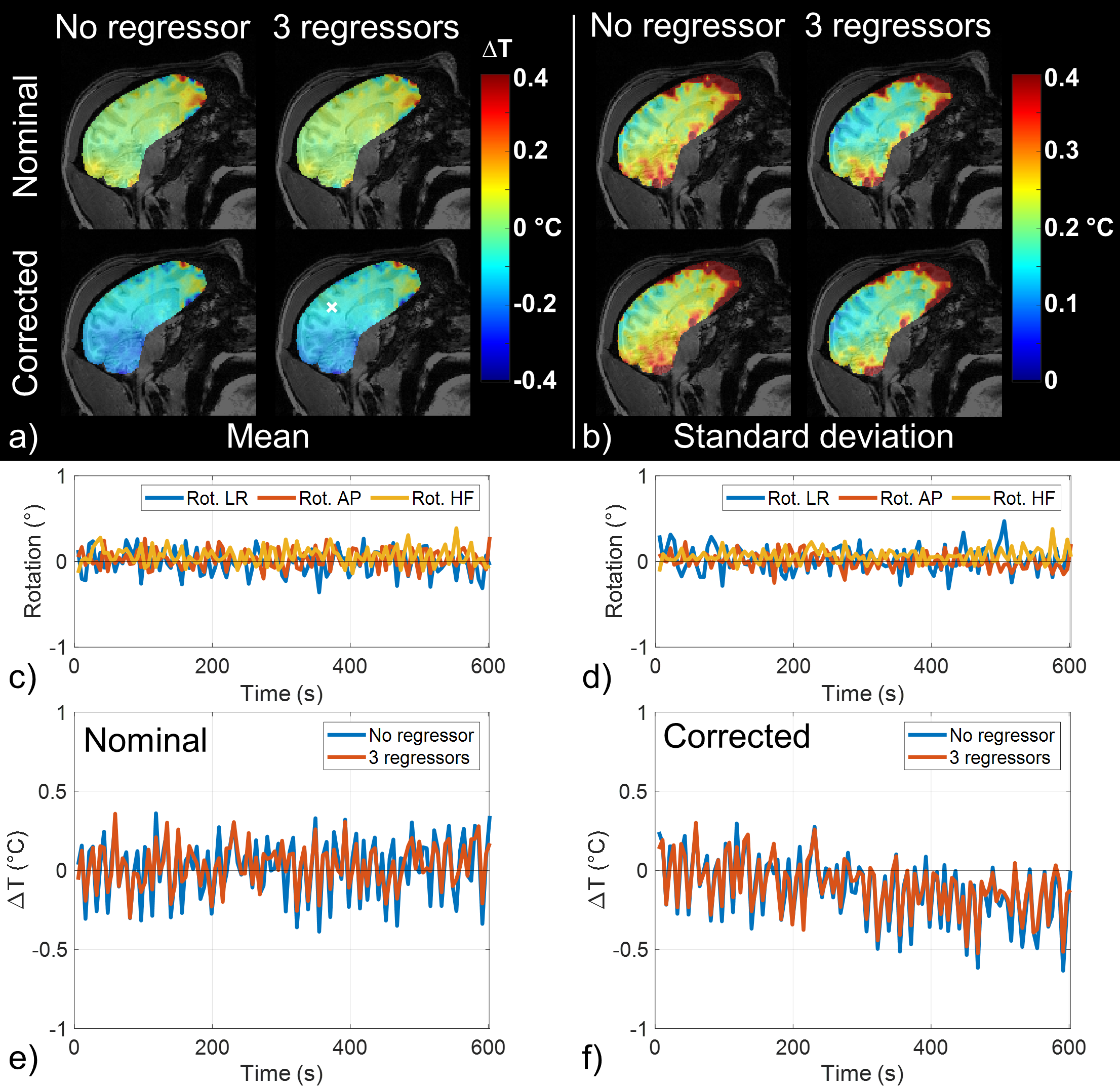
Figure 2: Scan without RF heating results for the macaque (run 1). (a) Mean ΔT maps and (b) corresponding SD maps computed from voxel-wise detrended signals. ΔT maps were computed from images reconstructed with nominal trajectories (upper row) and first order spherical harmonics field correction (lower row). ΔT computed from phase images corrected with rotation regressors around LR, AP and HF axis are also displayed. (c,d) Rotation parameters and (e,f) time evolution of ΔT computed from nominal and FS-corrected images, respectively, in the voxel indicated by the white cross in (a).
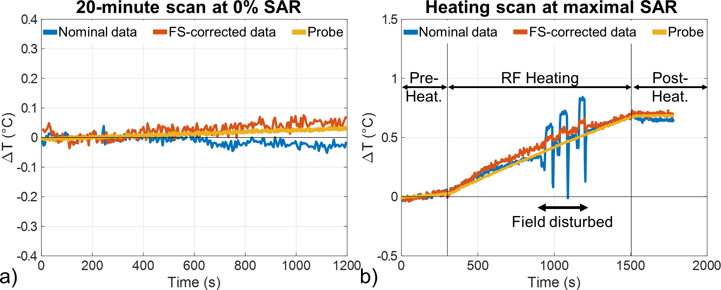
Figure 1: (a) Temperature variation measured in vitro with the fiber optical probe and from nominal and field sensor (FS) corrected MRT images at probe location over the 20 minutes scan at approximately 0% of the maximal SAR. (b) Temperature variation measured during the heating scan (5 min pre-heating at 0% SAR + 20 min heating at maximal SAR + 5 min post-heating at 0% SAR) by the fiber optical probe and from nominal and FS-corrected MRT images at probe location, in field disturbed conditions.