Waqas Majeed1, Axel J. Krafft2, Sunil Patil1, Henrik Odéen3, John Roberts3, Florian Maier2, Dennis L. Parker3, and Himanshu Bhat1
1Siemens Medical Solutions USA Inc., Malvern, PA, United States, 2Siemens Healthcare GmbH, Erlangen, Germany, 3Department of Radiology and Imaging Sciences, University of Utah, Salt Lake City, UT, United States
1Siemens Medical Solutions USA Inc., Malvern, PA, United States, 2Siemens Healthcare GmbH, Erlangen, Germany, 3Department of Radiology and Imaging Sciences, University of Utah, Salt Lake City, UT, United States
We demonstrate that high quality PRF thermometry can be achieved in the brain and prostate at 0.55T using a segmented EPI approach.
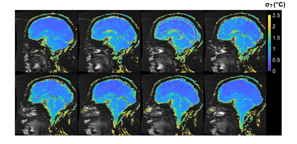
Figure 3: σT in the brain with ETL 49,
TE 81, 3.8s/frame: σT values of less than 2.5°C are overlaid on average magnitude image (8 out of 12 sagittal
slices). σT is less than 1°C in most of the brain, demonstrating excellent precision.
Average σT of 0.7 ± 0.2°C was observed in the ROI indicated on the figure. This protocol
offers a higher frame rate compared with that with ETL 33. The slight reduction
in σT, as compared with the protocol with ETL
33, can be attributed to the longer TE.
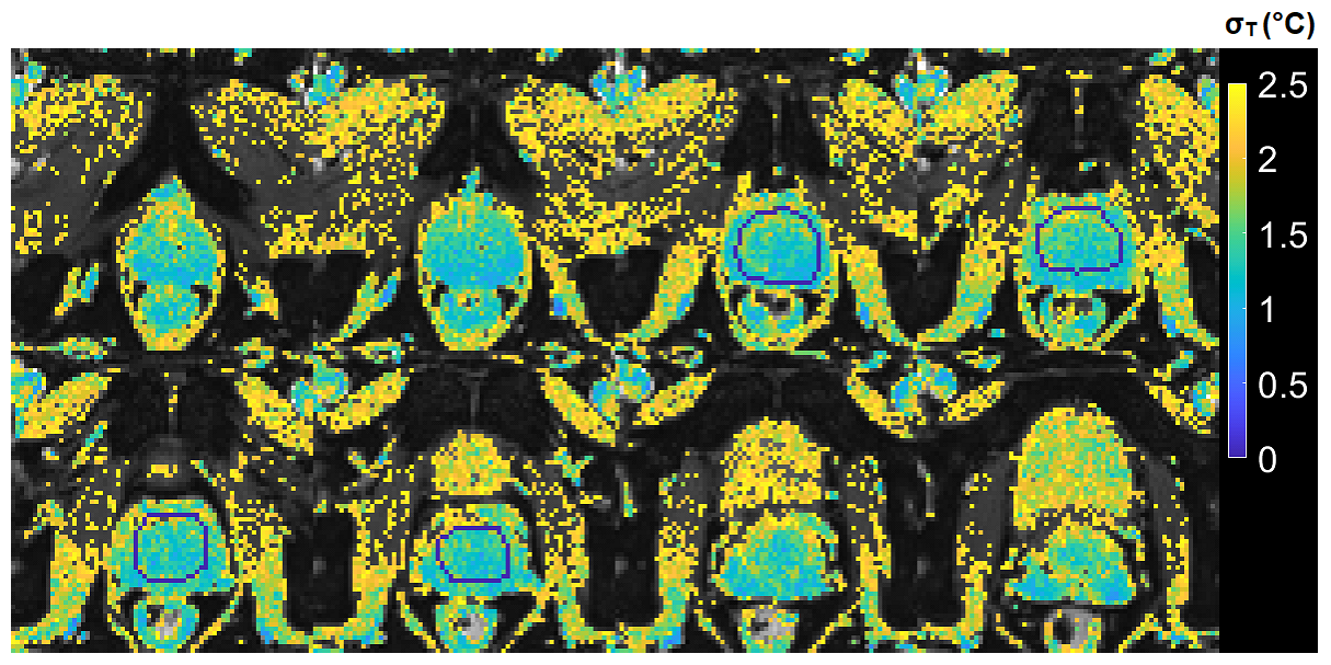
Figure 5: σT in the prostate with TR
100ms, TE 45ms, 7.2 s/frame. σT
values of less than 2.5°C are
overlaid on average magnitude image (8 out of 12 axial slices; cropped to show details). Average σT of
1.4±0.3°C was
observed in the ROI indicated on the figure, demonstrating good precision.