Chang-Sheng Mei1, Shenyan Zong2, and Guofeng Shen2
1Department of Physics, Soochow University, Taipei, Taiwan, 2Biomedical Instrument Institute, School of Biomedical Engineering, Shanghai Jiao Tong University, Shanghai, China
1Department of Physics, Soochow University, Taipei, Taiwan, 2Biomedical Instrument Institute, School of Biomedical Engineering, Shanghai Jiao Tong University, Shanghai, China
In this work, the conventional PRF method was evolved using the circle fitting for the temperature measurements in fat-containing tissues. As a result, the temperature accuracy by the proposed method in aqueous and adipose tissues was improved in our verification experiments.
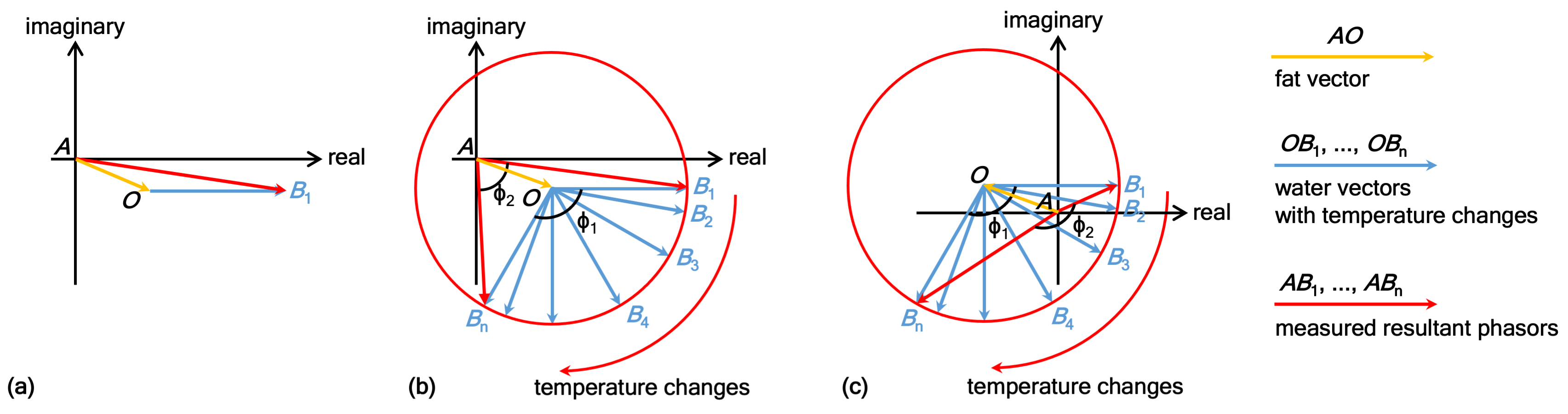
Fig. 1: The diagrams of water and fat phasors of MR data in Cartesian coordinates. In (a), the add-up phasor represents the actual phasor obtained from image, where the orange and blue arrows are phasors for fat and water protons, respectively. (b,c) show the water phase rotation around point 'O' caused by temperature changes and depict two scenarios of underestimating (φ2 < φ1) and overestimating (φ2 > φ1), respectively. A circle fitting can be applied here to determine the circle center 'O', which represents the fat phasor AO in (b) and (c), for getting the accurate water phase changes.
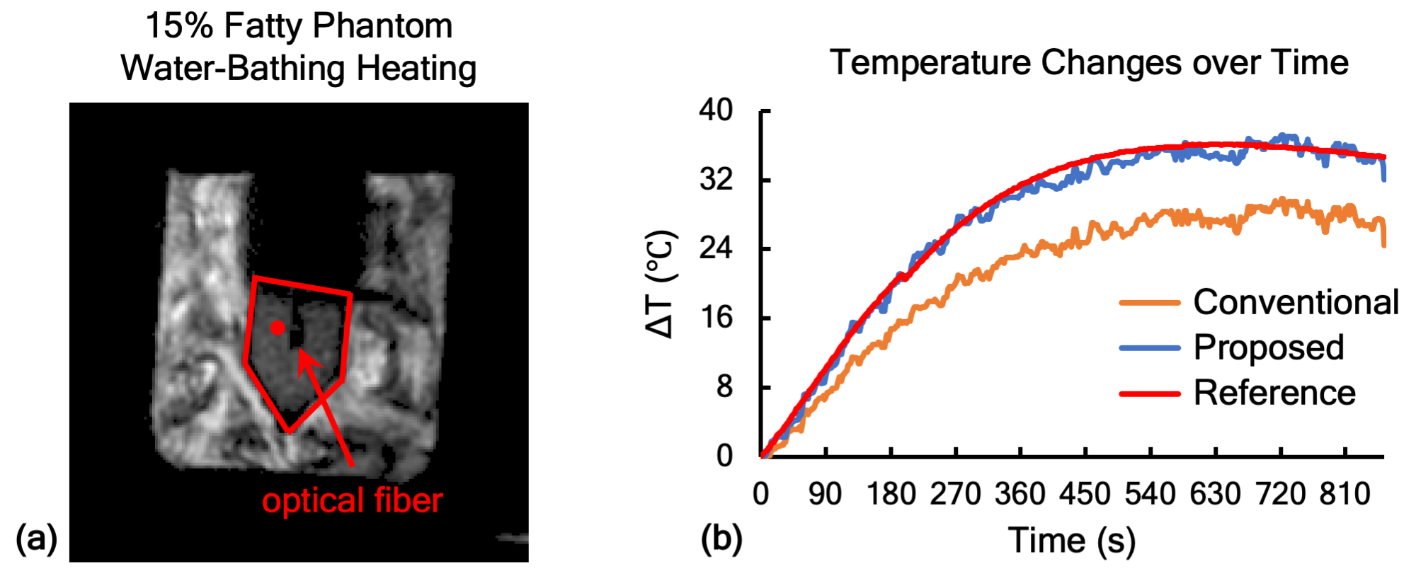
Fig. 3: Results of in vitro phantom experiments. In (a), the red pentagon and filled circle indicate the phantom region and the selected voxel for the plots of temperature evolution over time in (b). The orange, blue and red lines display the temperature changes obtained by the conventional method, the proposed method and the optical fiber. Approximately 33% of temperature error caused by 15% fat in phantom was observed and corrected in (b).