Tyler Blazey1, Galen D Reed2, Joel R Garbow1, and Cornelius von Morze1
1Mallinckrodt Institute of Radiology, Washington University in St. Louis, St. Louis, MO, United States, 2GE Healthcare, Dallas, TX, United States
1Mallinckrodt Institute of Radiology, Washington University in St. Louis, St. Louis, MO, United States, 2GE Healthcare, Dallas, TX, United States
We developed a metabolite-specific 2D EPI sequence with spatially-selective RF pulses for [1-13C]pyruvate and [1-13C]lactate at 4.7T. Using this sequence for in vivo imaging of hyperpolarized [1-13C]pyruvate resulted in improvements in temporal and spatial resolution compared to CSI.
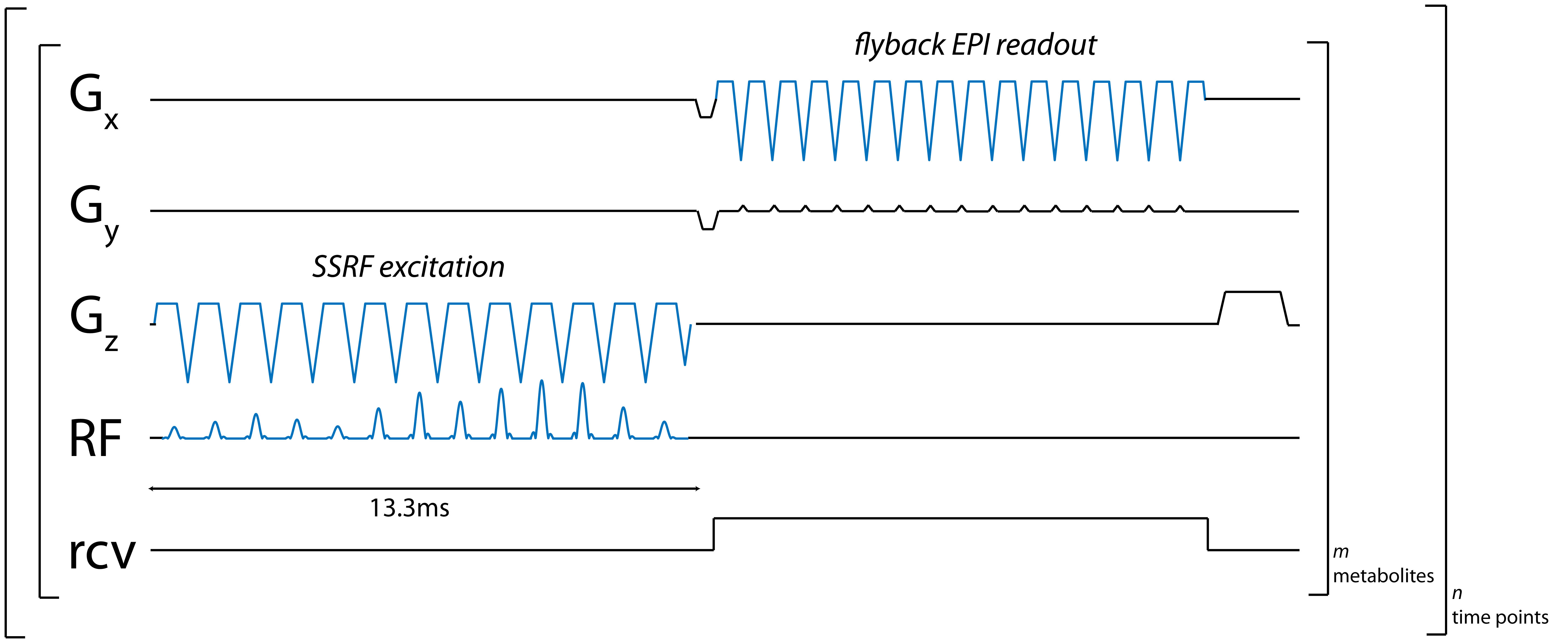
Figure 1: Pulse sequence diagram for metabolite-specific 2D EPI imaging of [1-13C]pyruvate and [1-13C]lactate at 4.7T. Lactate-only excitation pulse is shown.
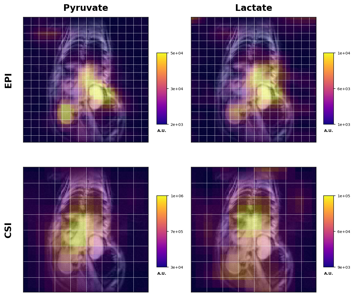
Figure 3: Metabolite images of [1-13C]pyruvate (1st column) and [1-13C]lactate (2nd column) in the liver and kidney of a rat following injection of [1-13C]pyruvate. Images in the first row were acquired using metabolite-specific EPI (Figure 1) while images in the second row were acquired using a standard CSI sequence. All images were obtained by summing signal over all time points. White lines are acquisition grids for each image. Images are shown with a 2x interpolation factor.