Dominik Weidlich1, Julius Honecker2, Claudine Seeliger2, Daniela Junker1, Marcus R. Makowski1, Hans Hauner2, Dimitrios C. Karampinos1, and Stefan Ruschke1
1Department of Diagnostic and Interventional Radiology, School of Medicine, Technical University of Munich, Munich, Germany, 2Else Kröner Fresenius Center for Nutritional Medicine, Technical University of Munich, Freising, Germany
1Department of Diagnostic and Interventional Radiology, School of Medicine, Technical University of Munich, Munich, Germany, 2Else Kröner Fresenius Center for Nutritional Medicine, Technical University of Munich, Freising, Germany
First feasibility of
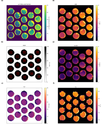
imaging-based ω-3 fraction mapping was demonstrated in
phantoms, as well as in vitro and in vivo in human adipose
tissue. In the phantom experiment, promising correlations were obtained between
imaging-based parameters and chromatography–mass spectrometry.

Figure
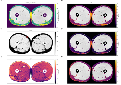
5: Resulting parameter maps of the thigh from the in vivo experiment: a) ω-3
fatty acid fraction, b) PDFF, c) T2*, d) ndb, e) nmidb and f) CL. A ROI
analysis (see ROI #1 in b)) yielded ω-3: 25.4±7.5%, ndb: 2.41±0.40,
nmidb: 0.67±0.26, CL: 18.41±0.54, T2*: 30.7±6.9ms. Low PDFF regions (e.g.
muscle tissue) were masked out in a) and e)-f).

