Chenyu He1, Xiaojun Guan2, Boliang Yu1, Hongjiang Wei3, and Yuyao Zhang1,4,5
1School of Information Science and Technology, ShanghaiTech University, Shanghai, China, 2Department of Radiology, The Second Affiliated Hospital, Zhejiang, University School of Medicine, Hangzhou, China, 3Institute for Medical Imaging Technology, School of Biomedical Engineering, Shanghai, Jiao Tong University, Shanghai, China, 4Shanghai Engineering Research Center of Intelligent Vision and Imaging, ShanghaiTech University, Shanghai, China, 5iHuman Institute, ShanghaiTech University, Shanghai, China
1School of Information Science and Technology, ShanghaiTech University, Shanghai, China, 2Department of Radiology, The Second Affiliated Hospital, Zhejiang, University School of Medicine, Hangzhou, China, 3Institute for Medical Imaging Technology, School of Biomedical Engineering, Shanghai, Jiao Tong University, Shanghai, China, 4Shanghai Engineering Research Center of Intelligent Vision and Imaging, ShanghaiTech University, Shanghai, China, 5iHuman Institute, ShanghaiTech University, Shanghai, China
Caudate nucleus (CN), putamen (Pu), ventral
pallidum (VP), globus pallidus (GP, GPi &
GPe), nucleus accumbens (NAC), subthalamic nucleus (STN), substantia nigra (SN, SNc & SNr) and red nucleus (RN) are preciously depicted in the MNI space in our work.
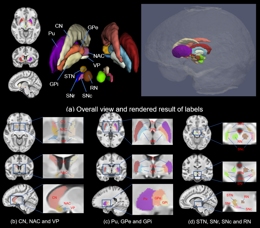
Visualization of hand-crafted nuclei depictions.
(a) Overall visualization of the parcellation map in axial, sagittal and
coronal views along with a 3D rendered view (label information only given on
right hemisphere); (b) Labeled CN, NAC and VP on the sections of three views;
(c) Labeled Pu, GPe and GPi on the sections of three views; (d) Labeled STN,
RN, SNr and SNc on the sections of three views.
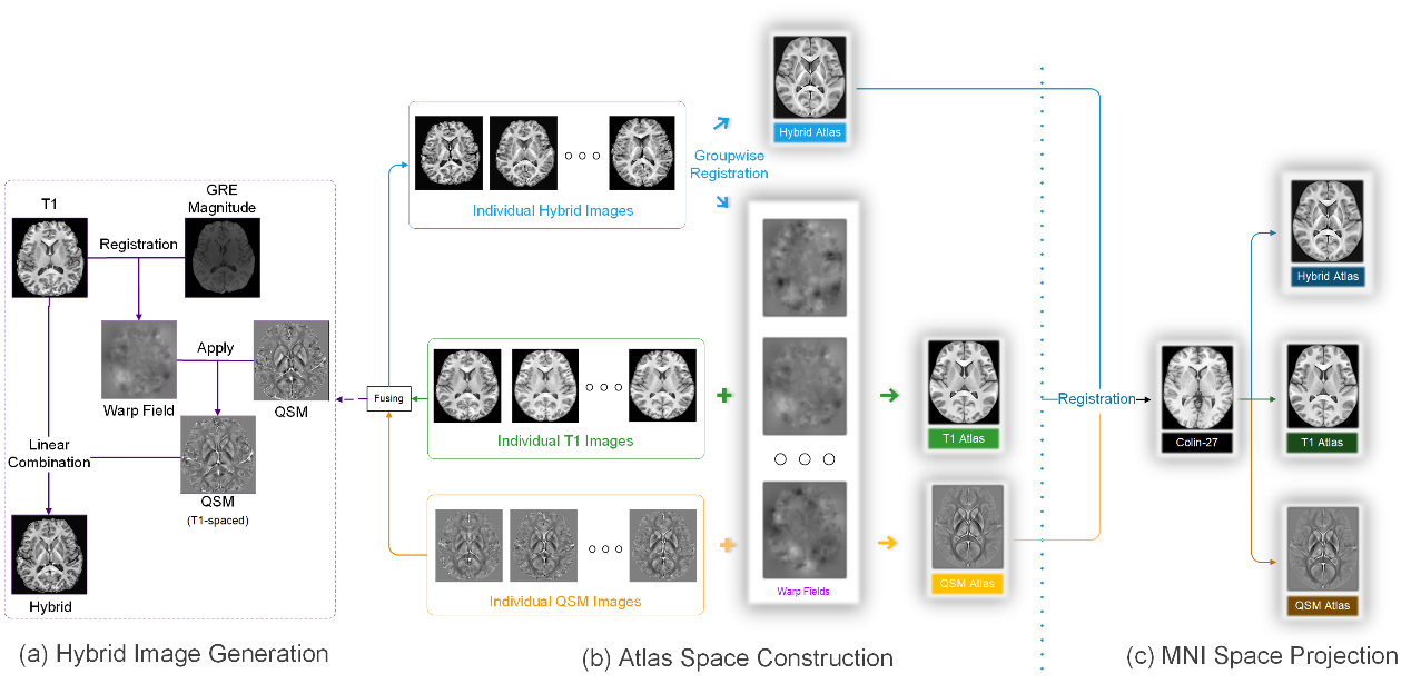
Pipeline of MNI space QSM atlas generation. (a)
Special normalization for individual T1w and QSM image; and hybrid image
generation. (b) Atlas space construction. (c) Guiding by T1w template, QSM
atlas is projected into MNI space.