Cassandra M.J. Wannan1, Shane Tonissen2, Bruce Tonge3, Efstratios Skafidas 1,2, Christos Pantelis1,2, and Warda T. Syeda1
1Melbourne Neuropsychiatry Centre, The University of Melbourne, Parkville, Australia, 2Department of Electrical and Electronic Engineering, The University of Melbourne, Parkville, Australia, 3Psychiatry Monash Health, Faculty of Medicine, Nursing and Health Sciences School of Clinical Sciences, Monash University, Melbourne, Australia
1Melbourne Neuropsychiatry Centre, The University of Melbourne, Parkville, Australia, 2Department of Electrical and Electronic Engineering, The University of Melbourne, Parkville, Australia, 3Psychiatry Monash Health, Faculty of Medicine, Nursing and Health Sciences School of Clinical Sciences, Monash University, Melbourne, Australia
- A multi-contrast image registration technique leverages information from multi-sequence MRI to non-linearly register an image to a reference space.
- The proposed technique outperforms single-contrast registration methods in a sample of 6-months old healthy infants.

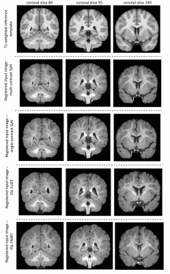
Figure 2: Exemplar skull-stripped T1w template images (top row) and corresponding single subject coronal slices from images registered using multi-contrast registration (second row), single-contrast ANTs (third row), FLIRT (fourth row) and FNIRT (last row). Single- and Multi-contrast images match template neuro-anatomy more closely in skull-stripped data compared to other methods.

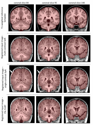
Figure 1: Exemplar T1w template coronal images in a single subject (top row) to visually compare multi-contrast registration (second row), single-contrast ANTs (third row) and FLIRT (bottom row) in whole-head data (reference space). Overlay: T1w template brain mask. Multi-contrast technique shows improved overlap between template and registered images. White arrows show underestimation of brain surface in single-contrast ANTs.
