Hajime Yokota1, Takayuki Sakai2, Masami Yoneyama3, Yansong Zhao4, and Takashi Uno1
1Department of Diagnostic Radiology and Radiation Oncology, Chiba University, Chiba, Japan, 2Department of Radiology, Eastern Chiba Medical Center, Togane, Japan, 3Philips Japan, Tokyo, Japan, 4Philips Healthcare, Cleveland, OH, United States
1Department of Diagnostic Radiology and Radiation Oncology, Chiba University, Chiba, Japan, 2Department of Radiology, Eastern Chiba Medical Center, Togane, Japan, 3Philips Japan, Tokyo, Japan, 4Philips Healthcare, Cleveland, OH, United States
MIXTURE provided high-resolution morphology images and T2 map simultaneously and can measure T2 values with high reproducibility for the cranial nerve, which was impossible in conventional multi-echo TSE and MIXTURE T2.
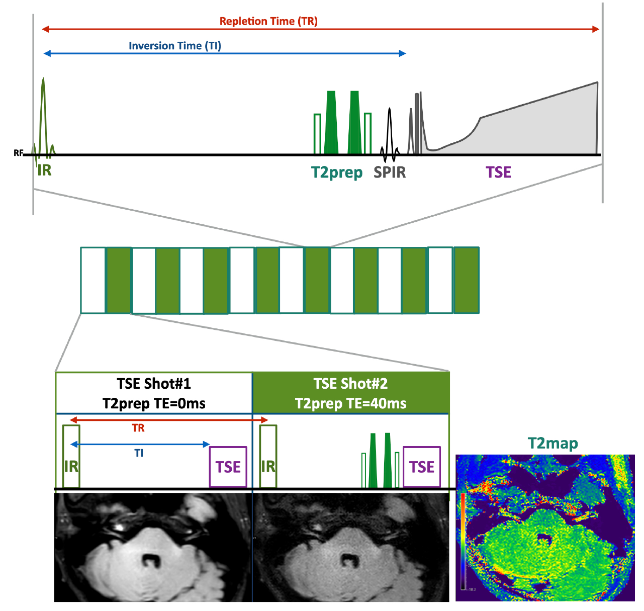
Figure 1. Scheme of the MIXTURE (Multi-Interleaved X-prepared tse with inTUitive RElaxometry)
T2-mapping was performed using T2-prepared 3D segmented turbo spin-echo (TSE) with variable refocusing pulse trains. Two images with different TE (TE = 0 and 50ms) were acquired with interleaved acquisition. To obtain FLAIR contrast, inversion time (TI) is adjusted to suppress cerebrospinal fluid.
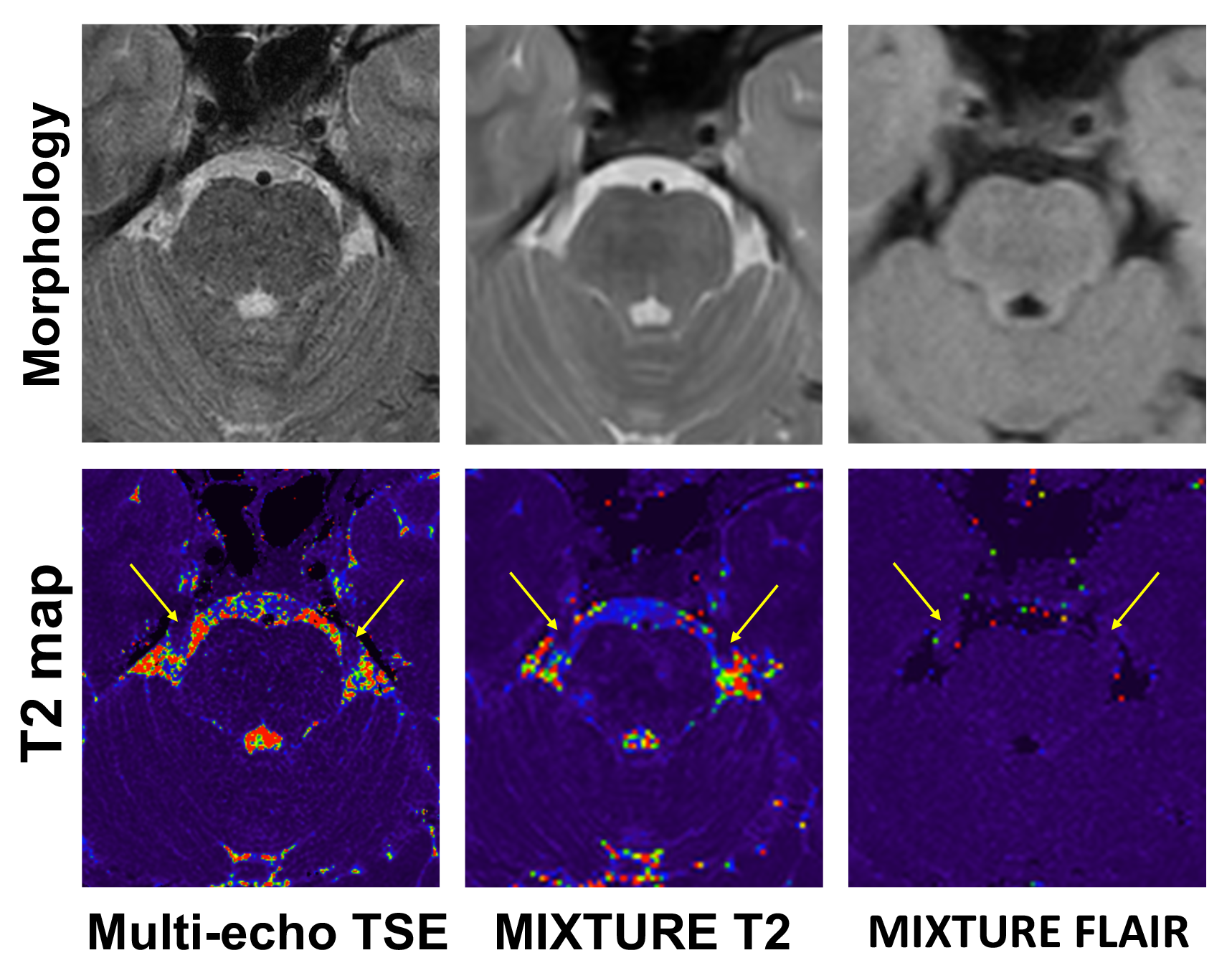
Figure 3. Trigeminal nerve presentation of multi-echo TSE, MIXURE T2, and MIXTURE FLAIR
Arrows indicates bilateral trigeminal nerves. The prepontine cistern around the trigeminal nerve looks noisy on T2 maps of multi-echo TSE and MIXURE T2, whereas the noise is reduced on the T2 map of MIXURE FLAIR.