Makoto Obara1, Yoshitomo Kikuchi2, Akio Hiwatashi2, Alexander Fischer 3, Yuta Akamine1, Tetsuo Ogino1, Masami Yoneyama1, Ronee Asad1, Yu Ueda1, Jihun Kwon1, and Marc Van Cauteren4
1Philips Japan, Tokyo, Japan, 2Departments of Clinical Radiology, Graduate School of Medical Sciences, Kyushu University, Fukuoka, Japan, 3Philips GmbH Innovative Technologies, Aachen, Germany, 4Philips Healthcare, Tokyo, Japan
1Philips Japan, Tokyo, Japan, 2Departments of Clinical Radiology, Graduate School of Medical Sciences, Kyushu University, Fukuoka, Japan, 3Philips GmbH Innovative Technologies, Aachen, Germany, 4Philips Healthcare, Tokyo, Japan
We developed deep learning
model for automatic brain metastasis detection using black- and bright-blood
images for input data. The results suggest the usefulness of using two
contrasts images as input, compared to single contrast input.
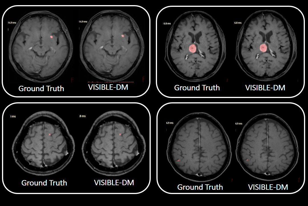
Figure 3: Four representative segmentation cases by VISIBLE-DM:
Segmented regions are shown in pink. Ground truth created by radiologists are also shown for comparison.
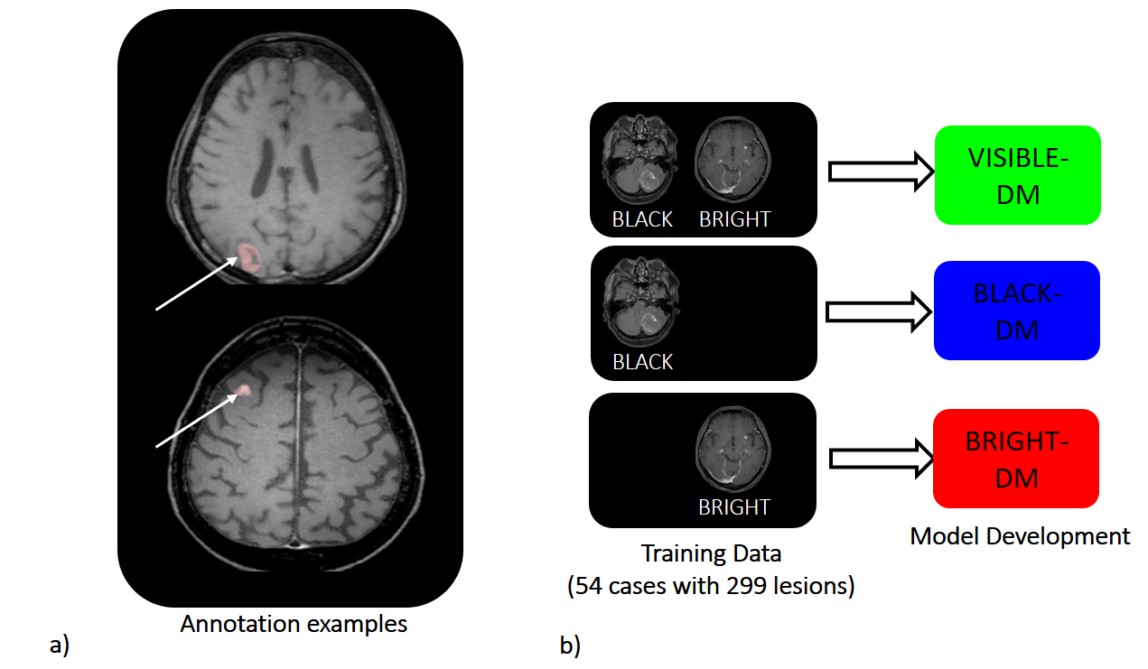
Figure 2: Model development: a) Annotation examples (white arrows), b) Models are
developed with three kinds of input data, black and bright, black only and
bright only images.