Rachael C Stickland1, Kristina M Zvolanek1,2, Stefano Moia3,4, Apoorva Ayyagari1,2, César Caballero-Gaudes3, and Molly G Bright1,2
1Physical Therapy and Human Movement Sciences, Feinberg School of Medicine, Northwestern University, Chicago, IL, United States, 2Biomedical Engineering, McCormick School of Engineering, Northwestern University, Evanston, IL, United States, 3Basque Center on Cognition, Brain and Language, Donostia, Spain, 4University of the Basque Country EHU/UPV, Donostia, Spain
1Physical Therapy and Human Movement Sciences, Feinberg School of Medicine, Northwestern University, Chicago, IL, United States, 2Biomedical Engineering, McCormick School of Engineering, Northwestern University, Evanston, IL, United States, 3Basque Center on Cognition, Brain and Language, Donostia, Spain, 4University of the Basque Country EHU/UPV, Donostia, Spain
Our results suggest that the relationship between baseline Cerebral Blood Flow and BOLD Cerebral Vascular Reactivity (CVR), across space and across people, is stronger when CVR is optimized for vascular delay and modelled with data including breathing tasks.
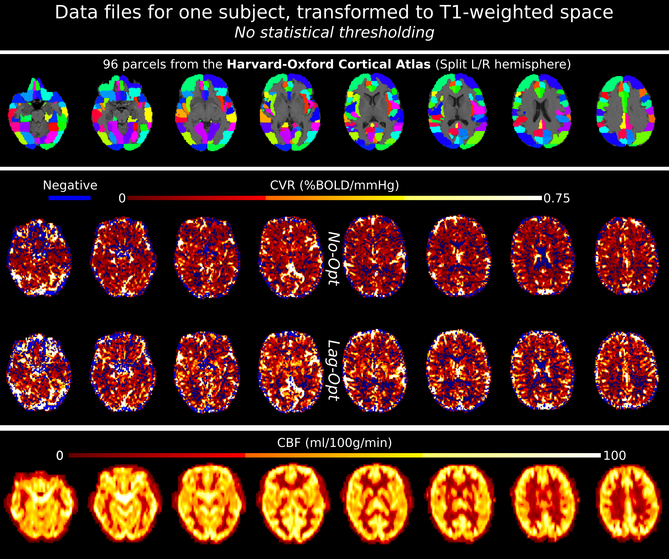
Example data files for one subject, in T1-weighted space. Top panel shows the Harvard-Oxford Cortical atlas: 48 regions split into left and right hemisphere parcels. The middle panel shows CVR maps, not optimized for lag (No-Opt) and optimized for lag (Lag-Opt), for the BH+REST data segment. The bottom panel shows baseline CBF maps. Average CBF and CVR were obtained from each parcel.
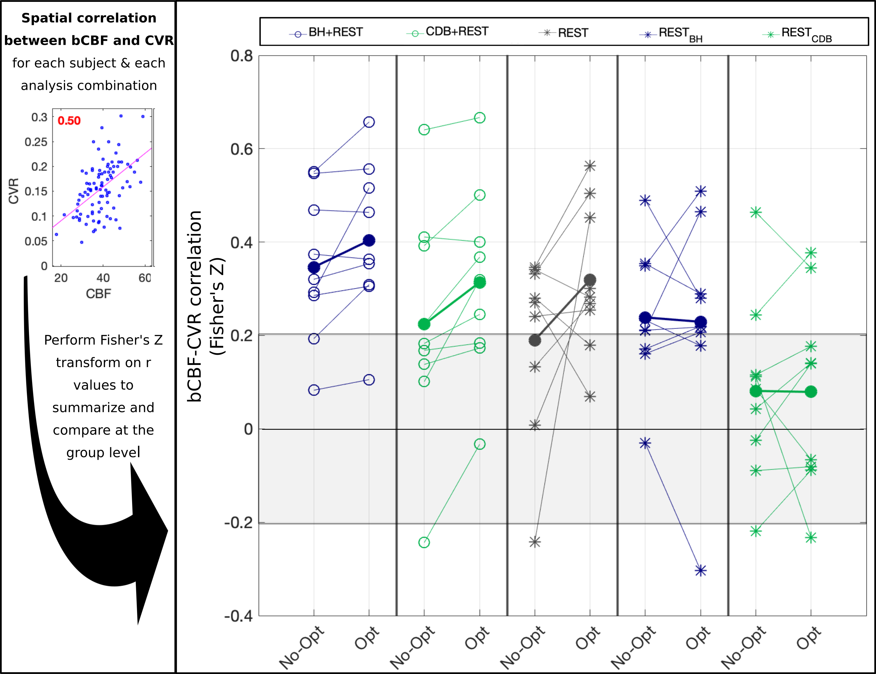
The association between bCBF and CVR values in 96 cortical parcels, compared across each of the five data segments (columns), for CVR values with no lag optimization (No-Opt) and CVR values with lag optimization (Opt). The thick lines that connect filled dots represent the group mean, and the thin lines represent each individual subject. The shaded grey box represents which correlations were not significant at the single subject level, based on a critical value of 1.96 (p<0.05, two-tailed).