Melanie Bauer1,2, Celine Berger1,2, Claudia Lenz1,2, Eva Scheurer1,2, and Christoph Birkl3
1Institute of Forensic Medicine, Biomedical Engineering, University of Basel, Basel, Switzerland, 2Institute of Forensic Medicine, Health Department Basel-Stadt, Basel, Switzerland, 3Department of Neuroradiology, Medical University of Innsbruck, Innsbruck, Austria
1Institute of Forensic Medicine, Biomedical Engineering, University of Basel, Basel, Switzerland, 2Institute of Forensic Medicine, Health Department Basel-Stadt, Basel, Switzerland, 3Department of Neuroradiology, Medical University of Innsbruck, Innsbruck, Austria
The orientation dependency of R2* is not influenced by the chosen
b-values for post mortem in situ and in vivo MRI scans.
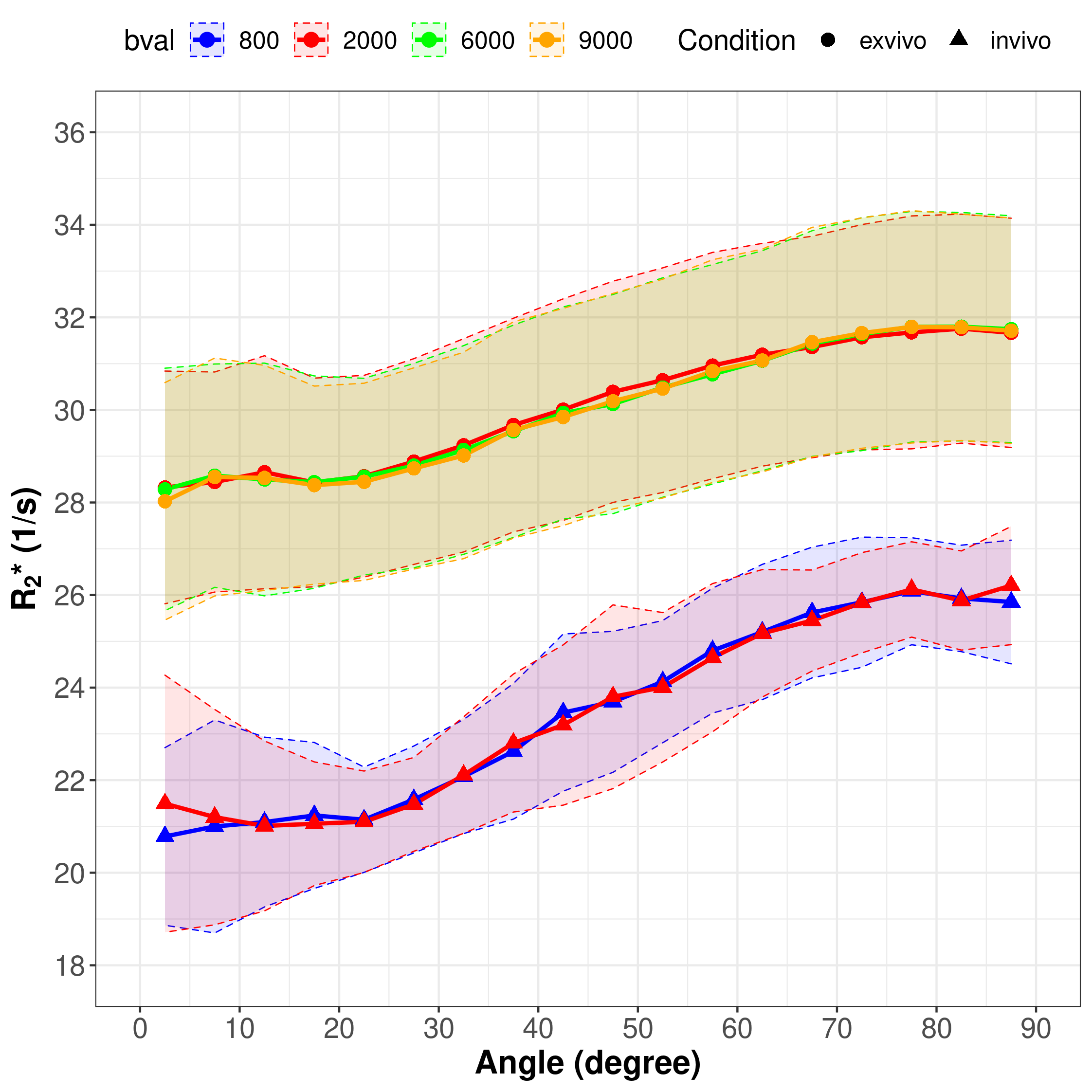
Figure 1: R2* behavior
in relation to increasing angle of white matter fibers. Post mortem data
recorded with b = 2000 s/mm2 (red), b = 6000 s/mm2 (green) and b = 9000 s/mm2 (yellow) are marked with dots and in vivo data
recorded with b = 800 s/mm2 (blue) and b = 2000 s/mm2 (red) are marked with triangles. The shaded
areas represent the 95% confidence interval of the respective data.
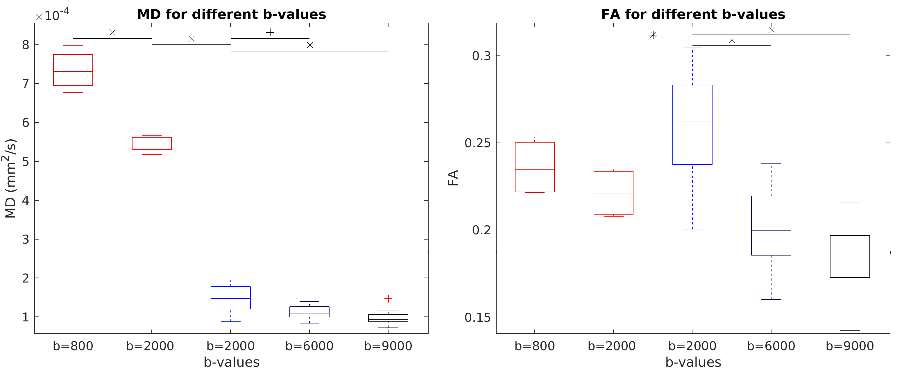
Figure 2: Boxplots
for MD (left) and FA (right) of different b-values used for in vivo (red) and
post mortem (blue) scans. Statistical significant differences < 0.05
are marked with *, < 0.01 with + and < 0.001 with x.