Alina Lopatina1,2, Stefan Ropele3, Renat Sibgatulin1, Jürgen R Reichenbach1,2,4, and Daniel Güllmar1
1Medical Physics Group / IDIR, Jena University Hospital, Jena, Germany, 2Michael-Stifel-Center for Data-Driven and Simulation Science, Jena, Germany, 3Department of Neurology, Medical University of Graz, Graz, Austria, 4Center of Medical Optics and Photonics Jena, Jena, Germany
1Medical Physics Group / IDIR, Jena University Hospital, Jena, Germany, 2Michael-Stifel-Center for Data-Driven and Simulation Science, Jena, Germany, 3Department of Neurology, Medical University of Graz, Graz, Austria, 4Center of Medical Optics and Photonics Jena, Jena, Germany
In this study, we proposed a method to translate susceptibility-weighted images of multiple sclerosis patients to healthy using generative adversarial networks. The method identified voxel information around the central veins and ventricles as MS-specific.
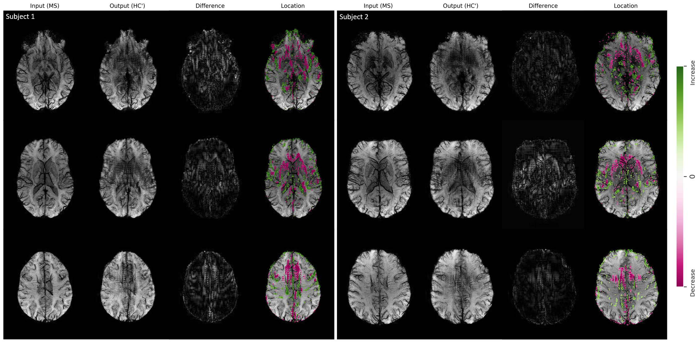
Figure
1. Image-to-image translation using FPG for two MS subjects for three slice
positions (in rows). The input images (MS) are translated to the healthy domain
(HC’). The absolute difference between the input and the translated images
reveals the disease-related voxels, which are overlaid on the original input
images. Green indicates an increase and pink a decrease in intensity.
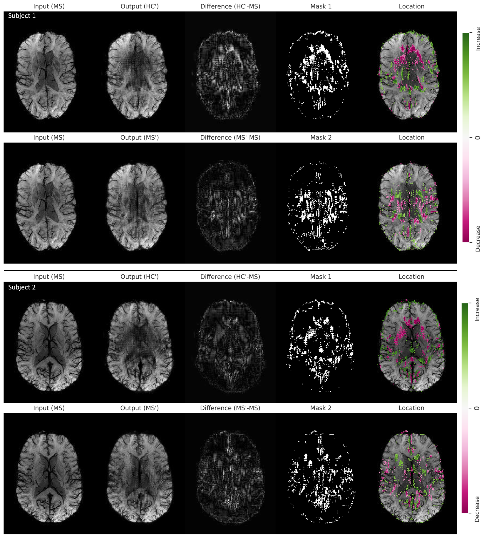
Figure
2. Image-to-image translation using FPG on two MS subjects for one slice
position. The input images (MS) are translated to the healthy domain (HC’)
(upper row) and to the diseased domain (MS’, lower row). The absolute
difference between the input and the translated images is threshold-limited to highlight
disease-related voxels, which are overlaid on the input images. Green indicates
an increase and pink a decrease in intensity.