Bhaswati Roy1, Sarah Choi2, Matthew J. Freeby 3, and Rajesh Kumar1,4,5,6
1Anesthesiology, University of California Los Angeles, Los Angeles, CA, United States, 2School of Nursing, University of California Los Angeles, Los Angeles, CA, United States, 3Medicine, Endocrinology - Diabetes and Metabolism, University of California Los Angeles, Los Angeles, CA, United States, 4Bioengineering, University of California Los Angeles, Los Angeles, CA, United States, 5Radiological Sciences, University of California Los Angeles, Los Angeles, CA, United States, 6Brain Research Institute, University of California Los Angeles, Los Angeles, CA, United States
1Anesthesiology, University of California Los Angeles, Los Angeles, CA, United States, 2School of Nursing, University of California Los Angeles, Los Angeles, CA, United States, 3Medicine, Endocrinology - Diabetes and Metabolism, University of California Los Angeles, Los Angeles, CA, United States, 4Bioengineering, University of California Los Angeles, Los Angeles, CA, United States, 5Radiological Sciences, University of California Los Angeles, Los Angeles, CA, United States, 6Brain Research Institute, University of California Los Angeles, Los Angeles, CA, United States
T2DM
patients show cognitive and mood changes, and brain tissue injury in those
regions; however, the underlying cause of tissue injury is unknown. We found
significantly reduced CBF in multiple brain areas and its associations with
functional deficits in those control sites in the condition.
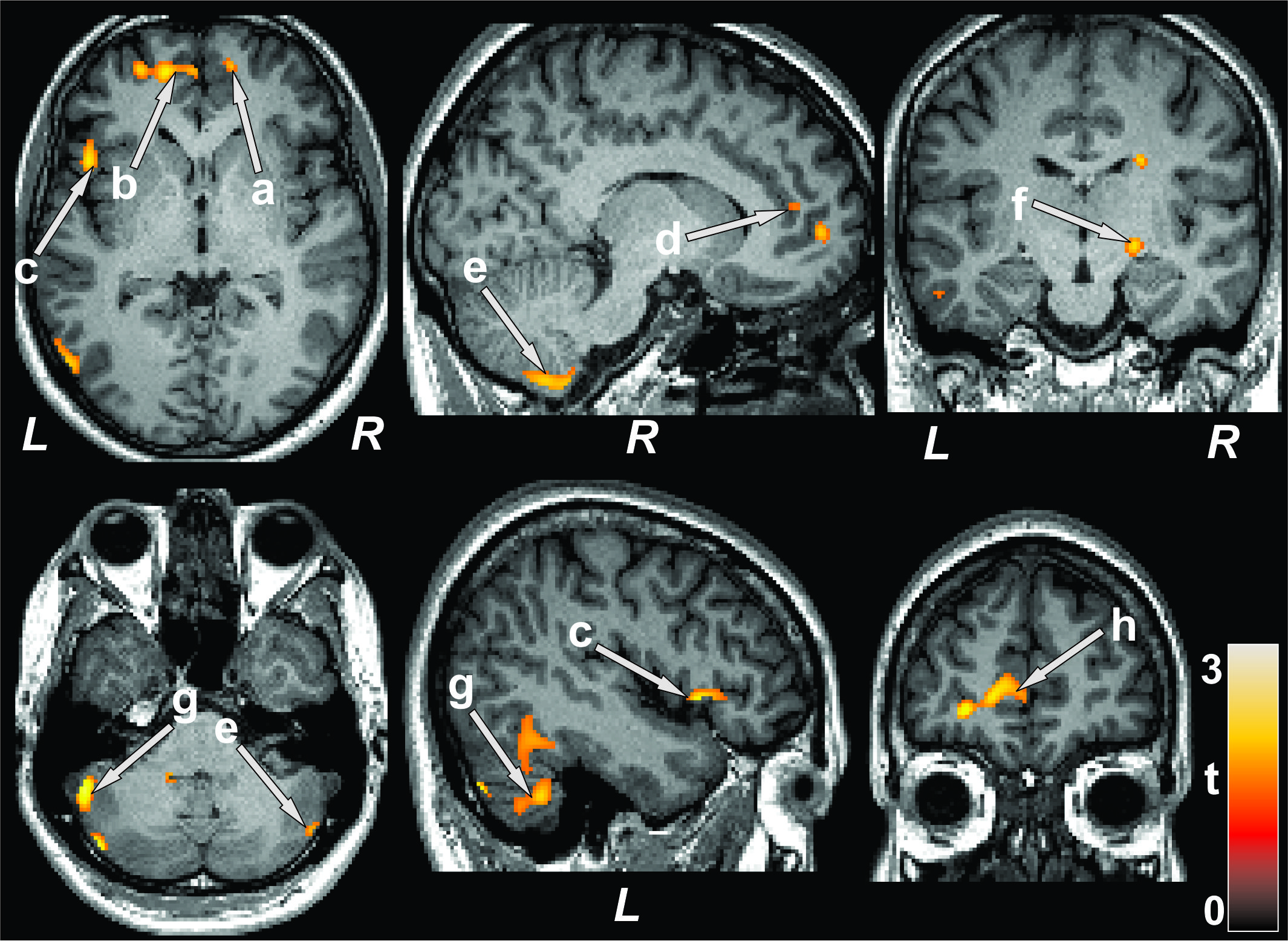
Figure 1: Brain regions with reduced cerebral blood flow in
T2DM patients over control subjects. The sites with reduced cerebral blood flow
in T2DM patients were included the bilateral prefrontal cortices (a, b), right
insula (c), bilateral cingulate (d, h), bilateral cerebellum (e, g), and right
thalamus (f). All images are in neurological convention (L = left; R = right).
Color bar indicates t-statistic values.
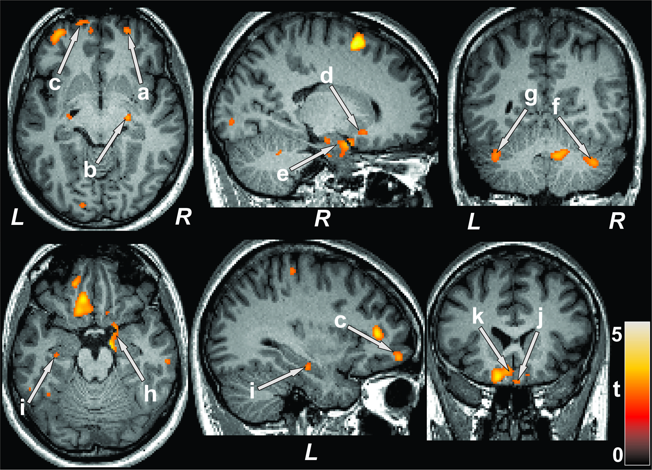
Figure 3: Cognition showed positive associations with cerebral
blood flow in T2DM patients. Positive correlations appeared between MoCA scores
and cerebral blood flow of the bilateral prefrontal cortices (a, c), right
thalamus (b), right putamen (d), bilateral hippocampus (e, i), bilateral
cerebellum (f, g), right amygdala (h), and bilateral basal forebrain (j, k).
Figure conventions are same as in Figure 1.