Fedel Machado-Rivas1,2, Lina Acosta Buitrago3, Jungwhan J Choi1,2, Onur Afacan1,2, Clemente Velasco-Annis1, Simon K Warfield1,2, Ali Gholipour1,2, and Camilo Jaimes1,2
1Radiology, Boston Children's Hospital, Boston, MA, United States, 2Radiology, Harvard Medical School, Boston, MA, United States, 3Universidad del Rosario, Bogota, Colombia
1Radiology, Boston Children's Hospital, Boston, MA, United States, 2Radiology, Harvard Medical School, Boston, MA, United States, 3Universidad del Rosario, Bogota, Colombia
Brain growth trajectories of fetal subjects with congenital diaphragmatic hernia (CDH) show global and localized volume loss when compared to typically developing fetuses. Volume differences between controls and CDH subjects are associated with hernia morphometric characteristics.
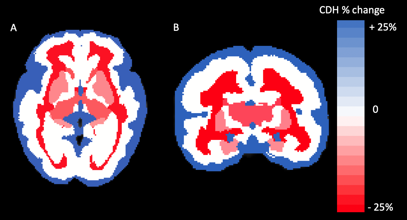
Axial (A) and corononal (B) heat map of segments differences between controls and CDH subjects. Percent change for CDH subjects when compared to controls is displayed. Segments in blue represent volume gain, while segments in red correspond to volume loss. White segments were not significantly different from controls.
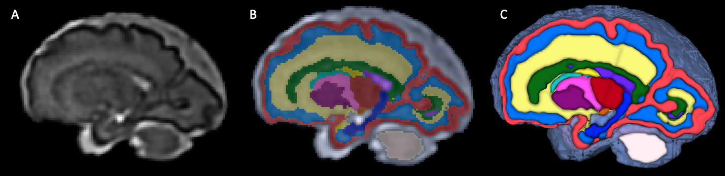
(A)T2-weighted super-resolution volume reconstruction with inter-slice motion correction, and intensity normalization of a T2-weighted fetal acquisition (29 week-old fetus). (B) Propagation of fetal atlas labels. (C) 3D volumetric rendering of propagated labels.