Celine Berger1,2, Claudia Lenz1,2, Melanie Bauer1,2, Eva Scheurer1,2, and Christoph Birkl3
1Institute of Forensic Medicine, Department of Biomedical Engineering, Basel, Switzerland, 2Institute of Forensic Medicine, Health Department Basel-Stadt, Basel, Switzerland, 3Department of Neuroradiology, Medical University of Innsbruck, Austria
1Institute of Forensic Medicine, Department of Biomedical Engineering, Basel, Switzerland, 2Institute of Forensic Medicine, Health Department Basel-Stadt, Basel, Switzerland, 3Department of Neuroradiology, Medical University of Innsbruck, Austria
R2* increased with increasing fiber angle in both post mortem in situ and in vivo, whereby a decreased R2* orientation dependency was observed post mortem compared to in vivo.
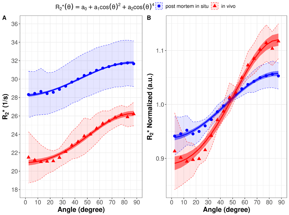
Figure 2: R2* as a function of the WM fiber angle averaged over the in vivo subjects (red) and the post mortem subjects (blue) fitted with the absolute values (A) and normalized to the global mean WM R2* (B). The shaded areas represent the 95% CI of the measured data.
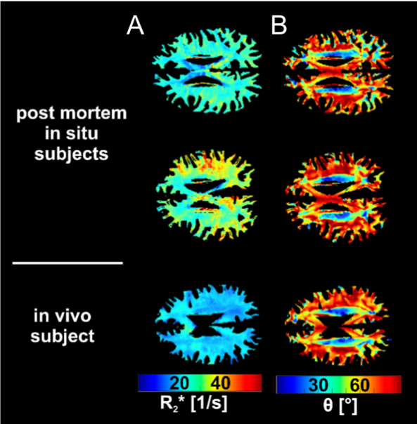
Figure 1: Representative maps of R2* (A) and fiber angle θ (B) of two post mortem subjects with brain temperatures of 5.6°C (top row) and 14.9 °C (middle row) and one in vivo subject shown in the bottom row.