Nikhil Deveshwar1,2 and Peder E. Z. Larson1,2
1Department of Radiology and Biomedical Imaging, University of California, San Francisco, San Francisco, CA, United States, 2UC Berkeley - UCSF Graduate Program in Bioengineering, Berkeley and San Francisco, CA, United States
1Department of Radiology and Biomedical Imaging, University of California, San Francisco, San Francisco, CA, United States, 2UC Berkeley - UCSF Graduate Program in Bioengineering, Berkeley and San Francisco, CA, United States
Ultrashort-T2* fraction parameter maps generated at different scans show similar looking structures in the same volunteer. The distributions of the ultrashort-T2* fractional component in various brain ROIs show similar distributions suggesting this technique is reproducible.
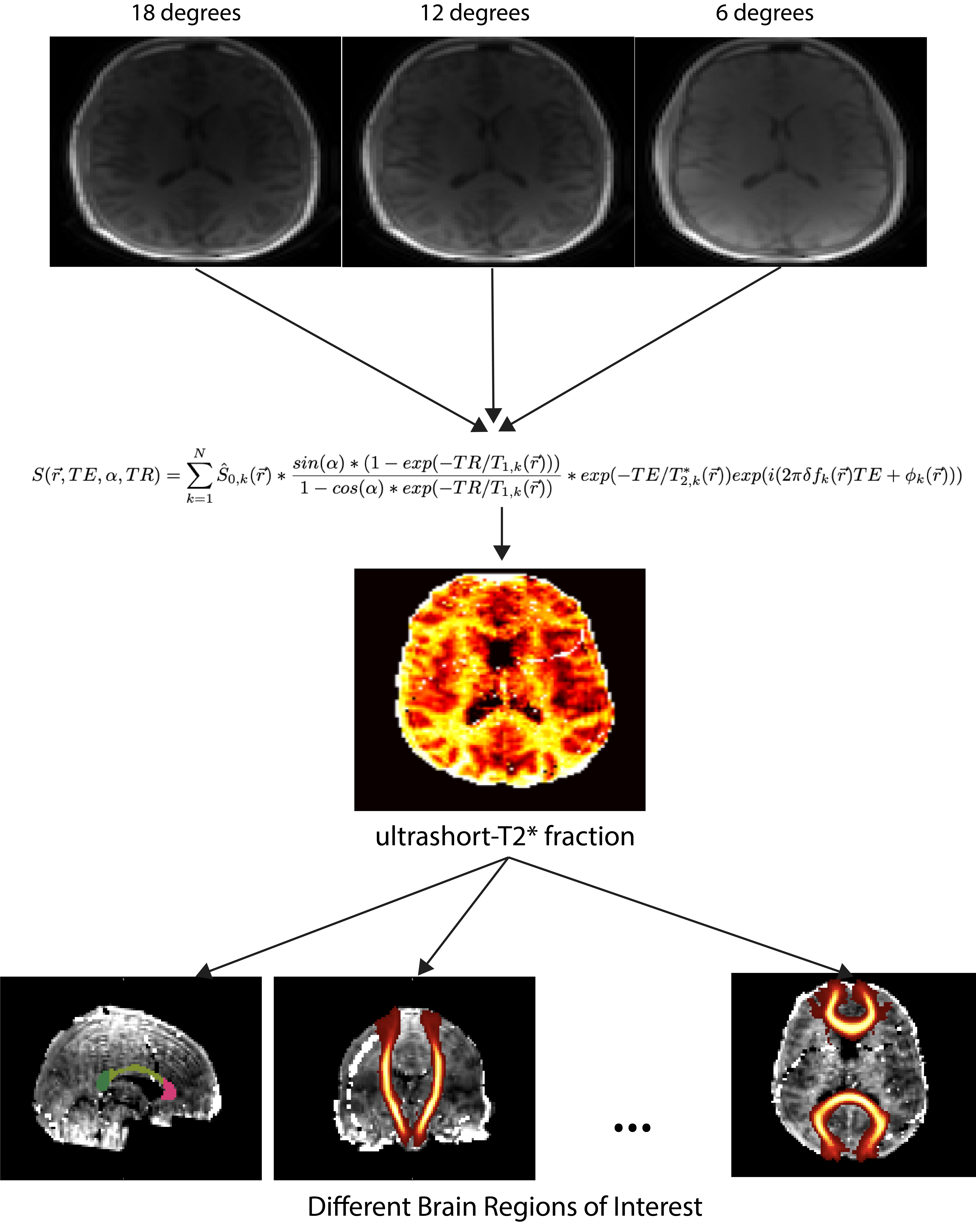
Figure 1: The proposed image processing pipeline. UTE scans at three different flip angles (18, 12, and 6 degrees) are fit with the following signal model. The resulting parameter map corresponding to the ultrashort-T2* fractional component is then used to isolate values in various brain ROIs.
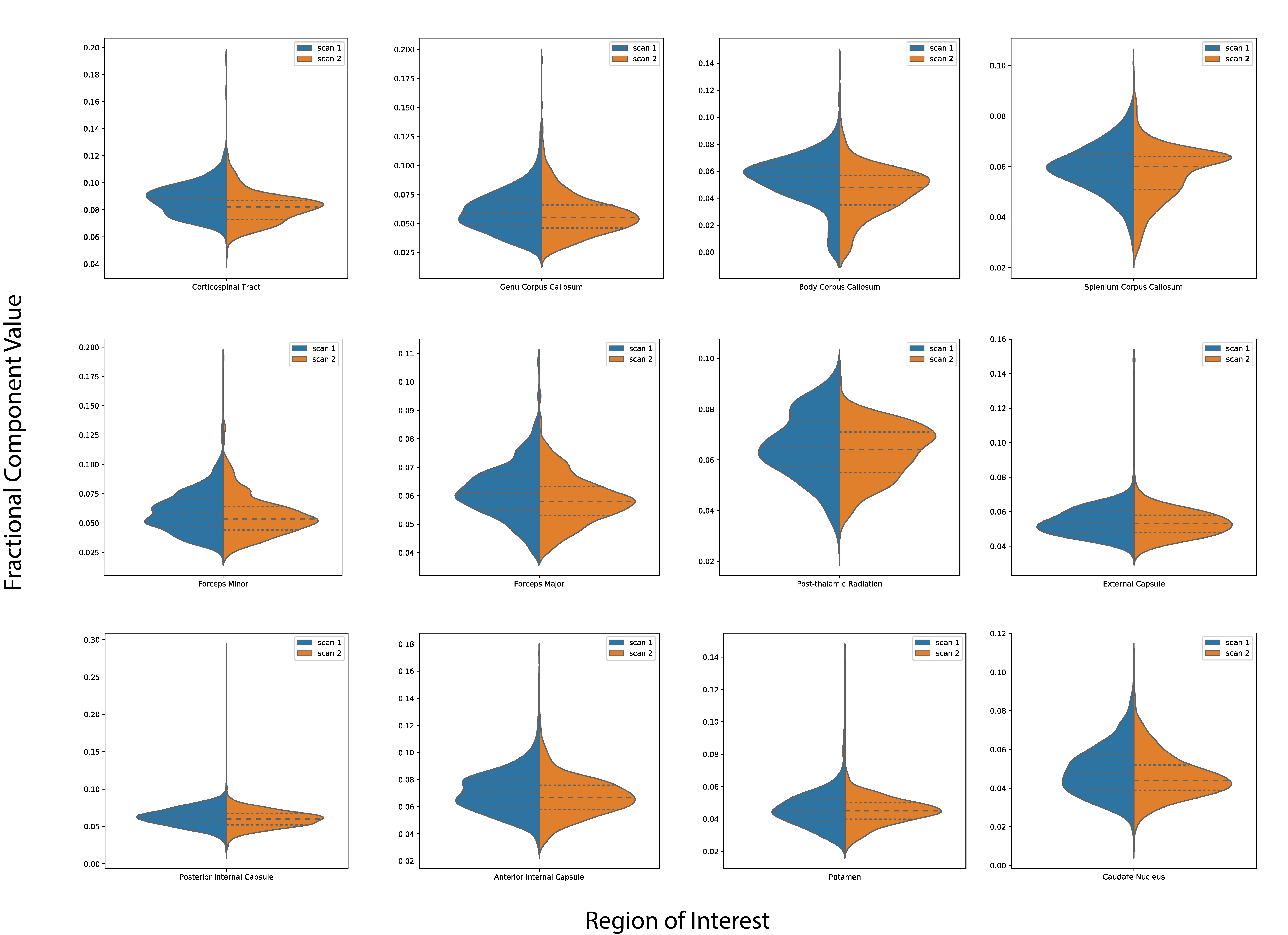
Figure 3: Split violin plots comparing the distributions of the ultrashort-T2* component fraction in various brain ROIs across 4 healthy volunteers. The dashed lines represent the interquartile range of each distribution.