Jodi Karlyn Watt1,2,3, Stefan Pszczolkowski1,2,3, Yue Xing1,2,3, Christopher Tench1,2,3, Dorothee Auer1,2,3, and Alzheimer's Disease Neuroimaging Initiative4
1Division of Clinical Neuroscience, University of Nottingham, Nottingham, United Kingdom, 2Sir Peter Mansfield Imaging Centre, University of Nottingham, Nottingham, United Kingdom, 3NIHR Nottingham Biomedical Research Centre, University of Nottingham, Nottingham, United Kingdom, 4Alzheimer's Disease Neuroimaging Initiative, Los Angeles, CA, United States
1Division of Clinical Neuroscience, University of Nottingham, Nottingham, United Kingdom, 2Sir Peter Mansfield Imaging Centre, University of Nottingham, Nottingham, United Kingdom, 3NIHR Nottingham Biomedical Research Centre, University of Nottingham, Nottingham, United Kingdom, 4Alzheimer's Disease Neuroimaging Initiative, Los Angeles, CA, United States
The relationship between CBF and tests of global cognition remains unknown. Principal Component Analysis was used to investigate the MoCA/cerebral perfusion relationship, providing preliminary support for a perfusion pattern which partially explains old-age cognitive decline.
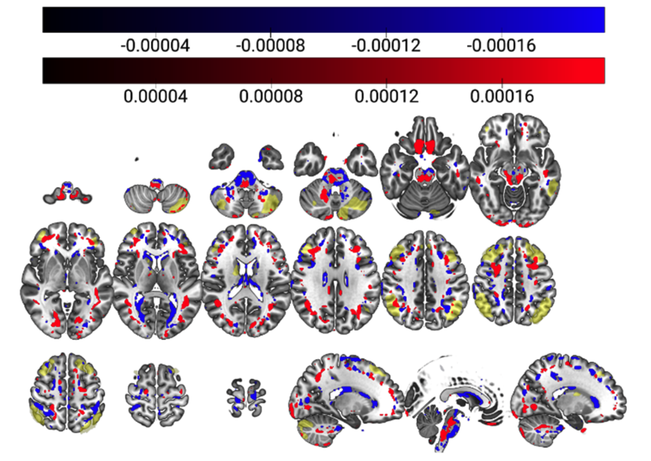
Figure 2: Combined surviving components (components 1, 4 and 42, 98% of r2 retained) overlaid on the MNI152, 2mm brain template. Negative associations with MoCA are depicted in blue, and positive in red, with the ECN map11 in yellow.
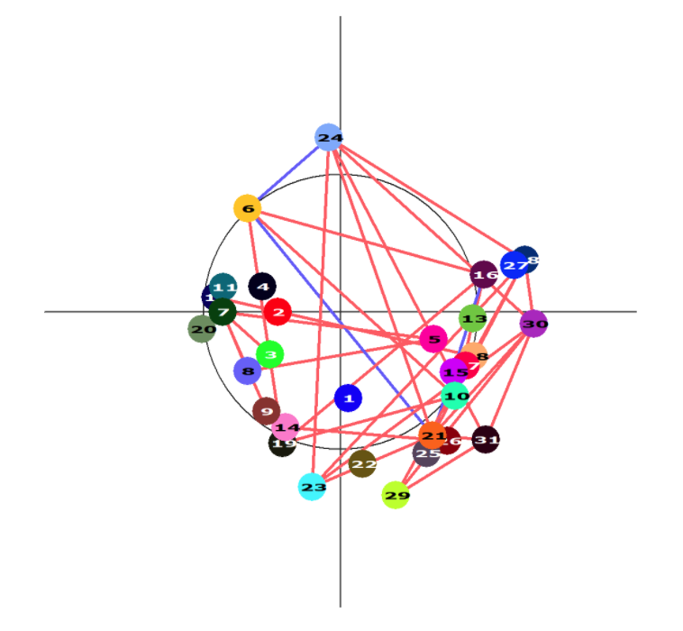
Figure 4: Colour-coded graph depicting the interrelatedness between any pairs of nodes. Positive associations are depicted with red edges, and negative with blue. The circle represents the mid-axial slice of the brain.