Cong Sun1, Aocai Yang1, Jiaguang Song2, Minhui Ouyang3, Jinxia Zhu4, Lei Xue5, Hao Huang3,6, and Gunagbin Wang1
1Radiology, Shandong Medical Imaging Research Institute, Cheeloo College of Medicine, Shandong University, Jinan, China, 2Ultrasound, Shandong Provincial Hospital Affiliated to Shandong University, Jinan, China, 3Radiology Research, Children’s Hospital of Philadelphia, Philadelphia, PA, United States, 4MR Collaboration, Healthcare Siemens Ltd., Beijing, China, 5MR Application, Siemens Healthineers Ltd., Jinan, China, 6Radiology, Perelman School of Medicine, University of Pennsylvania, Philadelphia, PA, United States
1Radiology, Shandong Medical Imaging Research Institute, Cheeloo College of Medicine, Shandong University, Jinan, China, 2Ultrasound, Shandong Provincial Hospital Affiliated to Shandong University, Jinan, China, 3Radiology Research, Children’s Hospital of Philadelphia, Philadelphia, PA, United States, 4MR Collaboration, Healthcare Siemens Ltd., Beijing, China, 5MR Application, Siemens Healthineers Ltd., Jinan, China, 6Radiology, Perelman School of Medicine, University of Pennsylvania, Philadelphia, PA, United States
Fetuses
with various kinds of complex congenital heart disease had higher values of
venous blood oxygen saturation than did normal fetuses of the same gestational
age, as measured with quantitative susceptibility mapping in utero.
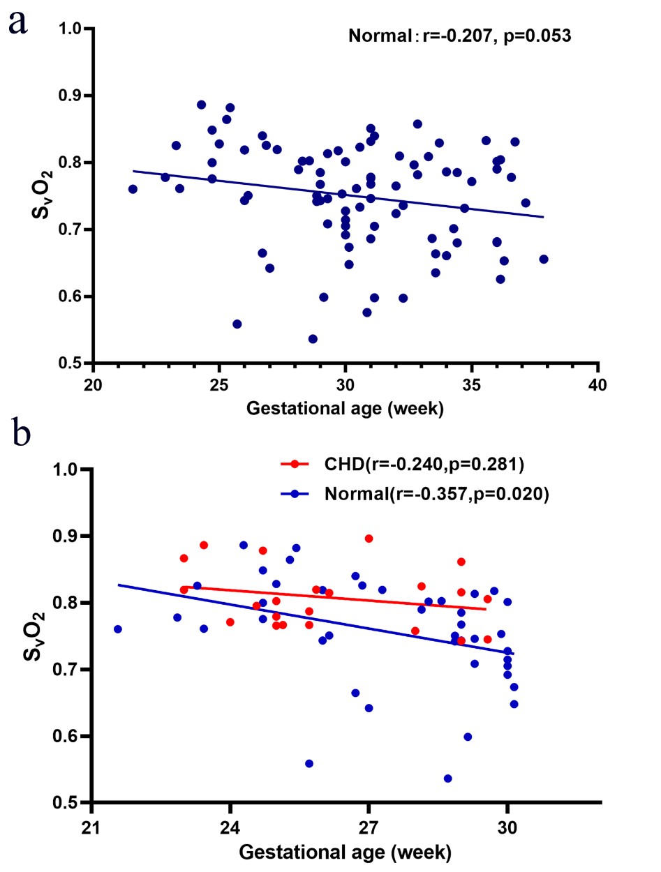
Figure
4a: The
venous blood oxygen saturation (SvO2)
alterations across gestational age (GA) of the normal group: SvO2 =
-0.004223*GA + 0.8783. Figure 4b: SvO2 alterations across gestational age between the
congenital heart disease (CHD) group (SvO2 = -0.00514*GA + 0.942) and
GA-matched normal group (SvO2 =
-0.01205*GA + 1.087) and their corresponding fitting curve. Red circles and red curves represent the CHD group; blue circles and blue curves represent the normal group.
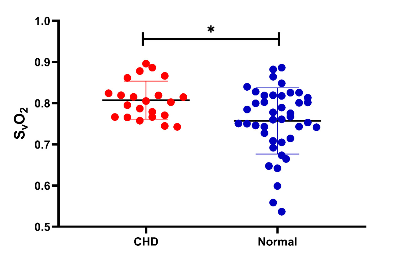
Figure 5: Venous blood oxygen
saturation (SvO2) values in the congenital heart disease (CHD) group and
gestational age (GA)-matched normal group, along with their corresponding mean and 95%
CI SvO2 values. In the analysis of covariance, the SvO2 value showed a significant difference between the CHD (80.8%±4.6%) group and the GA-matched
normal group (75.7%±8.0%) after the effects of GA were excluded (p=0.038).