Guangqi Li1, Xin Shao1, Xinyu Ye1, Xiaodong Ma2, and Hua Guo1
1Center for Biomedical Imaging Research, Department of Biomedical Engineering, School of Medicine, Tsinghua University, Beijing, China, 2Center for Magnetic Resonance Research, Radiology, Medical School, University of Minnesota, Minneapolis, MN, United States
1Center for Biomedical Imaging Research, Department of Biomedical Engineering, School of Medicine, Tsinghua University, Beijing, China, 2Center for Magnetic Resonance Research, Radiology, Medical School, University of Minnesota, Minneapolis, MN, United States
This
study demonstrates that single-shot spiral sampling can be adopted to acquire whole-brain diffusion tensor imaging
with shorter TE and higher SNR. In addition, the in vivo results show that single-shot
spiral DWI provides
accurate DTI metrics, and has high anatomical accuracy.
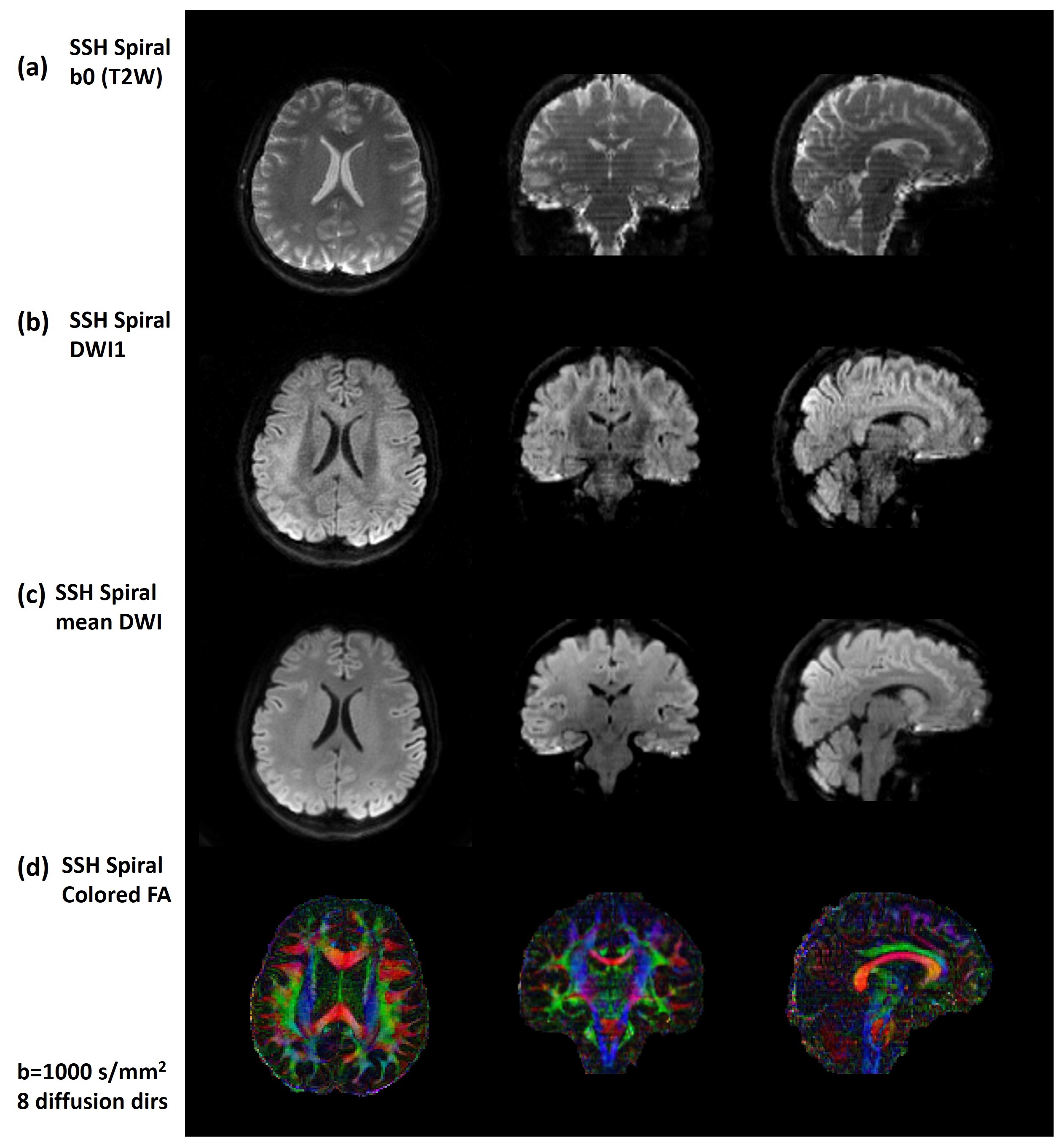
Figure 4: Axial,
coronal and sagittal planes of whole-brain diffusion weighted images with 1.3mm
isotropic resolution using single-shot spiral sampling. (a) b0 (T2W) images.
(b) single diffusion weighted images (DWI1). (c) mean DWI and (d) colored FA
maps.
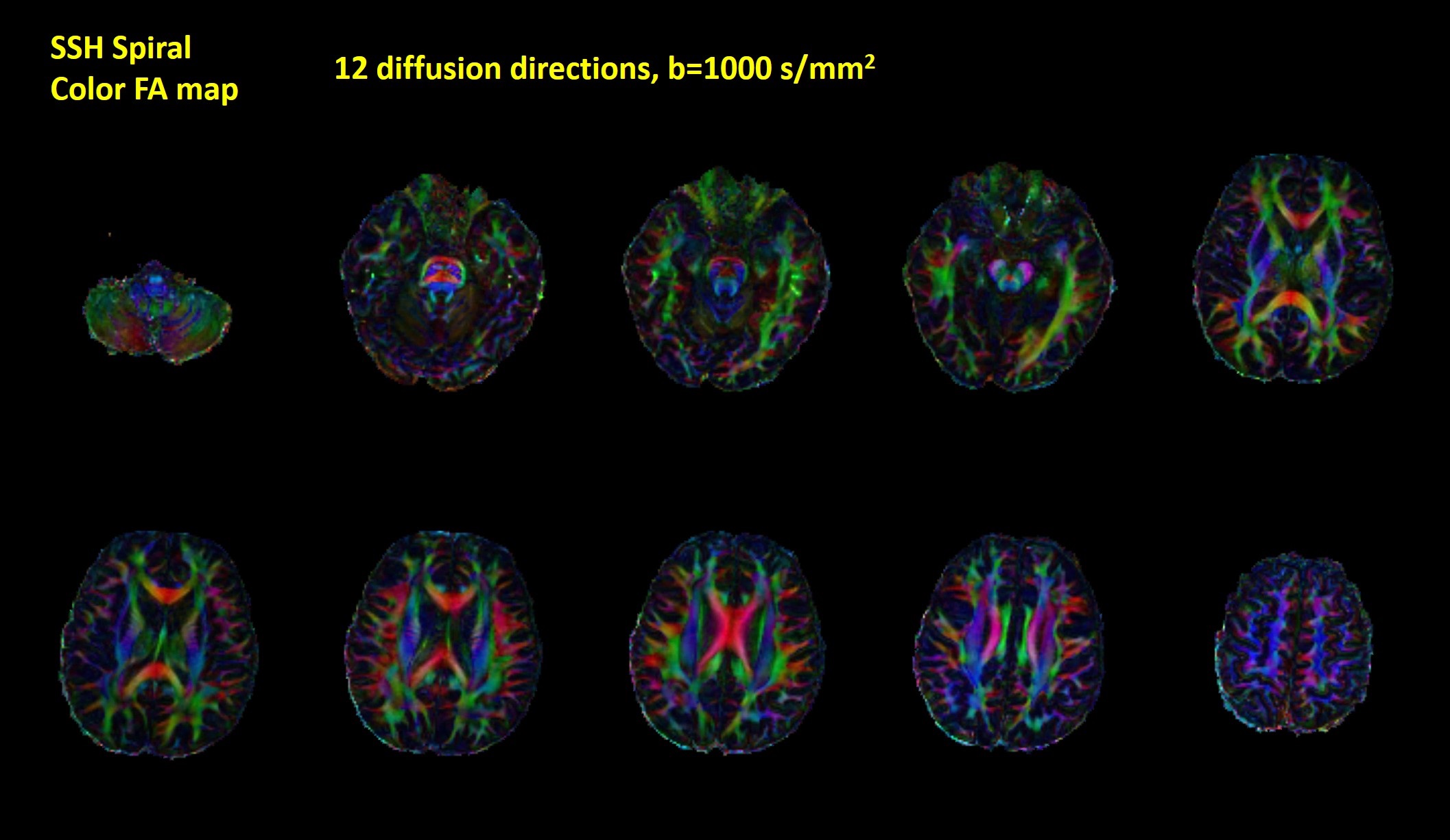
Figure
3: Colored FA maps of 10 representative
slices covering the whole brain. The results indicate that single-shot spiral
diffusion imaging can provide accurate DTI metrics.