Belinda Ding1, Iulius Dragonu2, Patrick Liebig3, Robin M Heidemann3, and Christopher T Rodgers1
1Wolfson Brain Imaging Centre, University of Cambrige, Cambridge, United Kingdom, 2Siemens Healthcare Limited, Firmley, United Kingdom, 3Siemens Healthineers, Erlangen, Germany
1Wolfson Brain Imaging Centre, University of Cambrige, Cambridge, United Kingdom, 2Siemens Healthcare Limited, Firmley, United Kingdom, 3Siemens Healthineers, Erlangen, Germany
This abstract showed that dynamic pTx pulses greatly reduces
signal dropouts in whole brain diffusion MRI at 7T when compared against
traditional circularly polarised pulses. This leads improvements in diffusion
tract definitions, especially in lower brain regions.
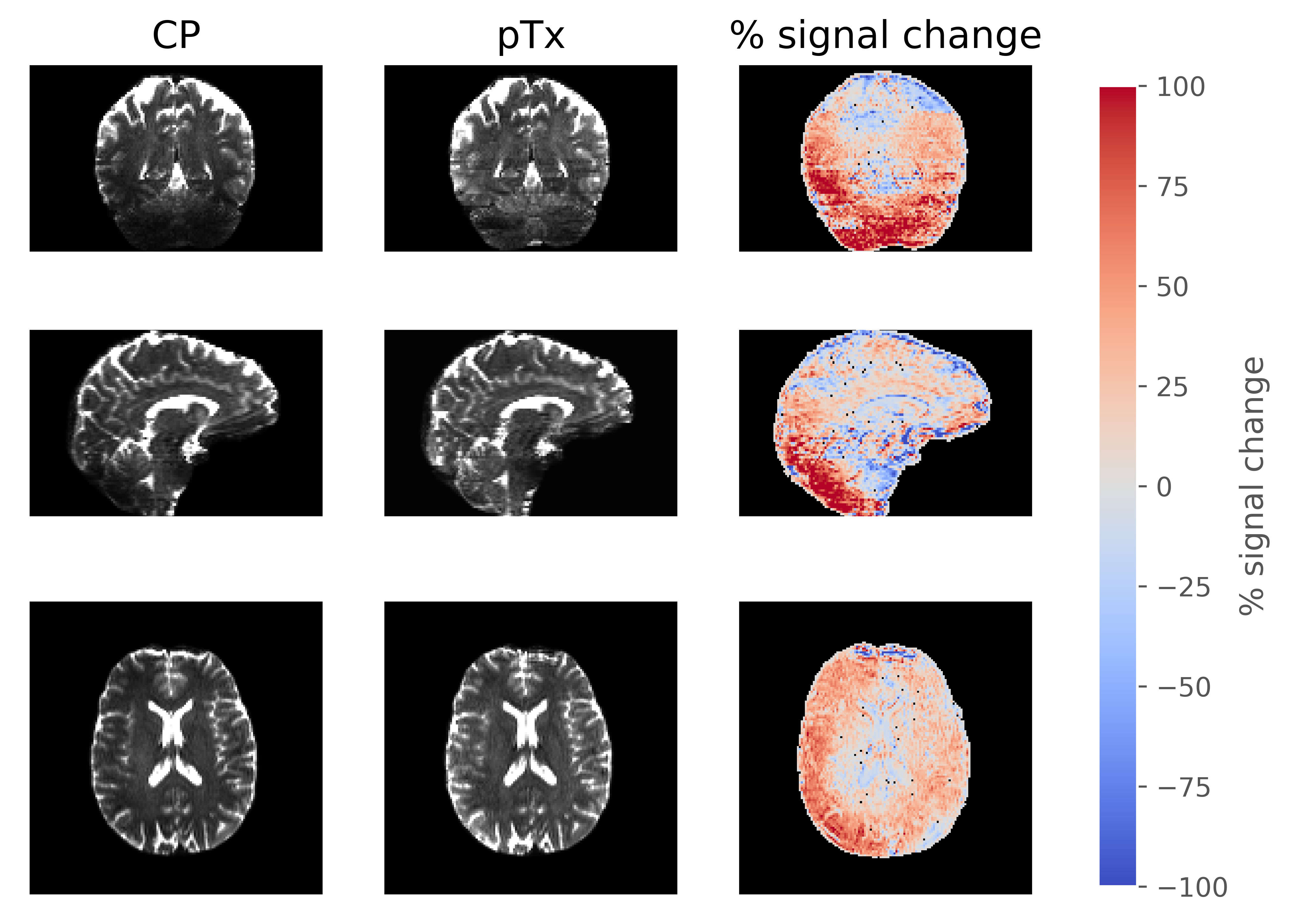
Figure 2: Direct comparison of the b=0 s/mm2
images between CP and pTx acquisition in three orientations. The right most
column shows the percentage signal change between the pTx and CP acquisition.
The signal change was calculated as 200% × [signal(pTx) – signal(CP)] /
[signal(pTx) + signal(CP)] .
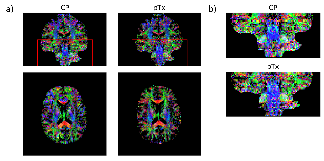
Figure 4: (a) Colour FA maps acquired with CP pulses
(left) and pTx spokes pulses (right). The maps show FA (in the range of [0.25
1]) with colours representing the orientation of the first eigenvector (red:
left-right; green: anterior-posterior; blue: inferior-superior). (b) Zoomed
region of interest as denoted by the red box in (a). Diffusion fibres are more
clearly seen in the pTx acquisition especially in the brainstem.