1Department of Radiology, Chiba University Hospital, Chiba, Japan, 2Diagnostic Radiology and Radiation Oncology, Graduate School of Medicine, Chiba University, Chiba, Japan, 3Philips Japan, Tokyo, Japan
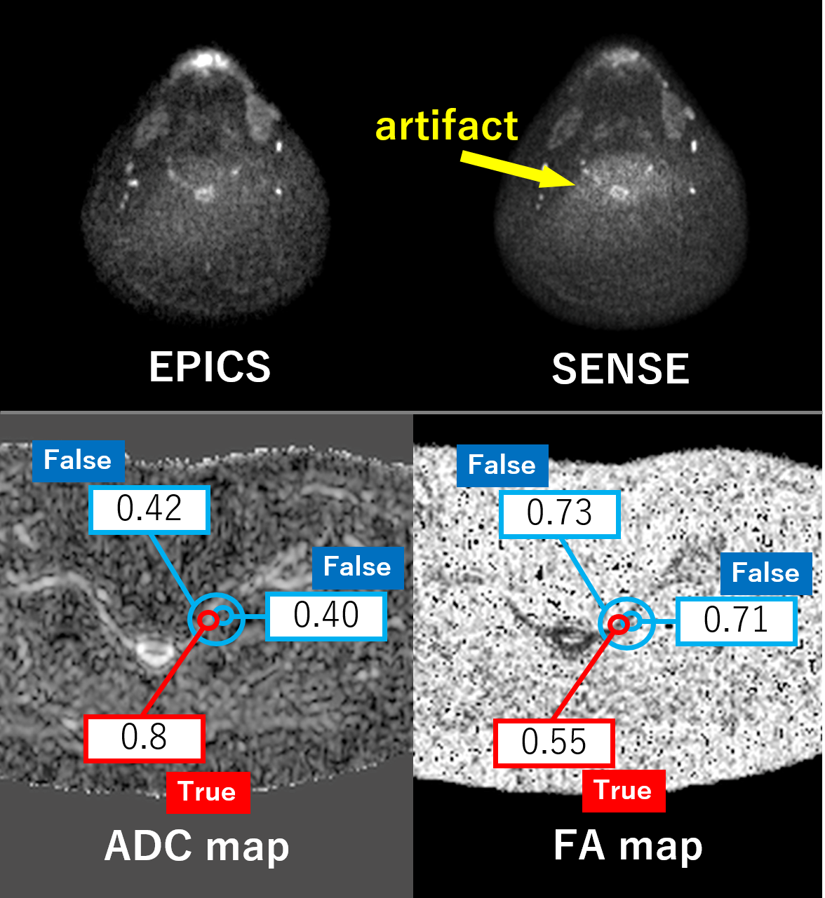
Figure 5. (Upper column) An example of noise-like artifacts in SENSE. The nerve root region is noisy, which may contribute to the reduced coefficients of variation in this region. (Lower column) There is a lot of noise in the area outside the nerve root. In the region of noise, ADC value is low and FA value is high. When the ROI is placed outside the nerve, the values are greatly shifted. It is important to draw ROIs on noise-free images.
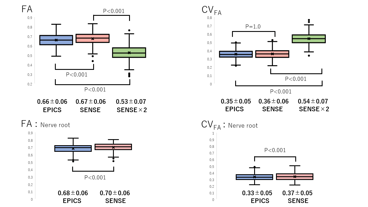
Figure 3.(Upper column) Fractional anisotropy (FA) and coefficient of variance (CVFA) of EPICS, SENSE, and SENSE×2. FA was significantly different among the three protocols, and CVFA was significantly smaller in EPICS and SENSE than in SENSE×2. (Lower column) Focusing on the nerve roots, FA in EPICS was significantly smaller and CVFA was smaller than SENSE.