Nora-Josefin Breutigam1, Daniel Christopher Hoinkiss1, Mareike Alicja Buck 1,2, Klaus Eickel1,2, Matthias Günther1,2, and David Porter3
1Imaging Physics, Fraunhofer MEVIS, Bremen, Germany, 2Faculty 01 (Physics/Electrical Engineering), University Bremen, Bremen, Germany, 3Imaging Centre of Excellence (ICE), University of Glasgow, Glasgow, Scotland
1Imaging Physics, Fraunhofer MEVIS, Bremen, Germany, 2Faculty 01 (Physics/Electrical Engineering), University Bremen, Bremen, Germany, 3Imaging Centre of Excellence (ICE), University of Glasgow, Glasgow, Scotland
Simultaneous
multi-contrast imaging (SMC) can be used to combine acquisition of
diffusion-weighted and T2*-weighted images into a single scan. Saturation effects can reduce
SNR and alter contrast. In this study, these effects are investigated in
simulations, in phantoms, and in vivo.
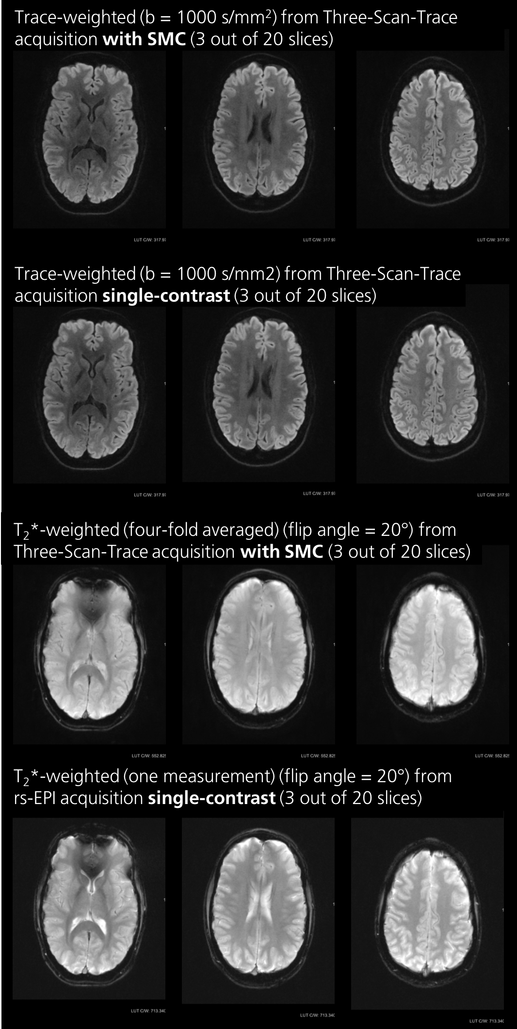
Figure 5: Data from a high-resolution, trace-weighted
acquisition with and without SMC. Three out of 20 slices are displayed. The potential
protocol for clinical use shows minimized SNR loss and contrast changes in the
DW images. The SNR loss in the T2*W case is higher, but this is more
than offset by four-fold averaging (from b-value of zero and three diffusion-gradient
directions with b = 1000 s/mm2). Contrast change in T2*W
case is mainly due to high CSF signal saturation.
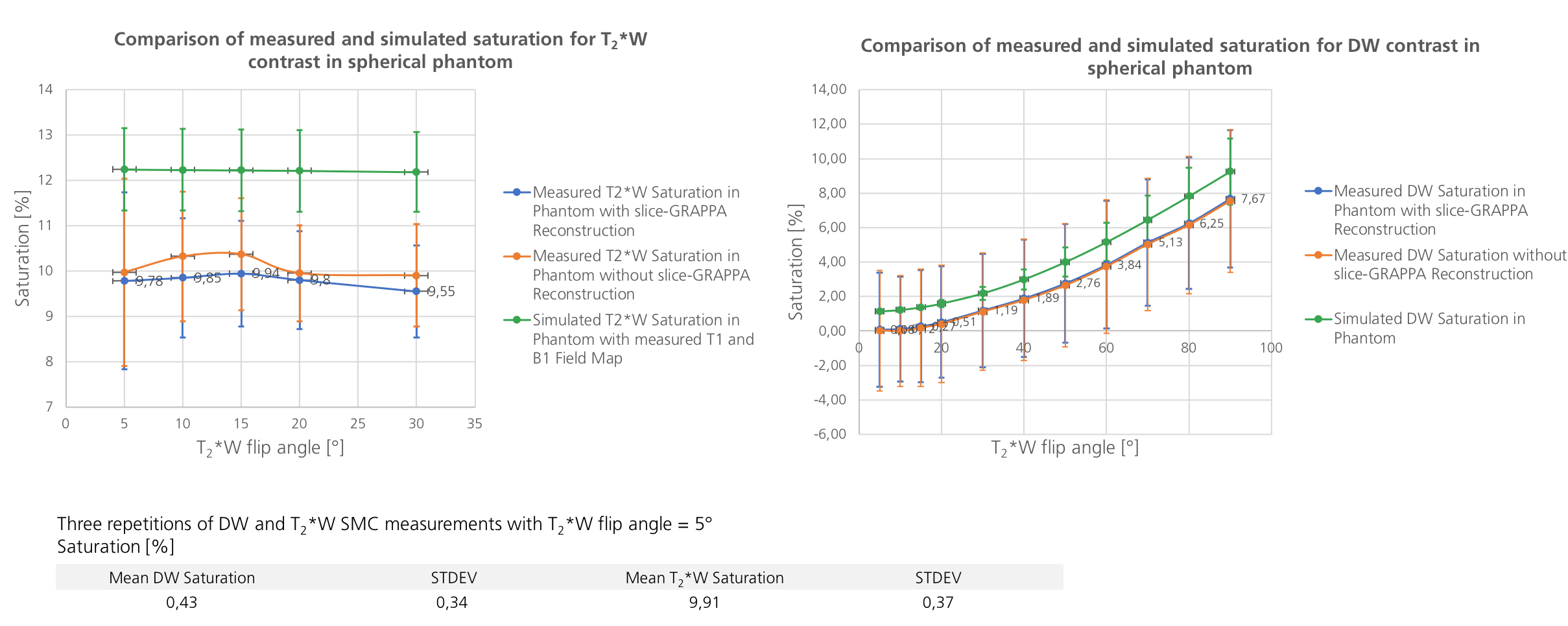
Figure 2: Measured and simulated
saturation for both contrast types in the phantom as a function of the T2*W
excitation flip angle with and without the use of split-slice GRAPPA during
image reconstruction. The trend of the three curves is almost identical. The
simulation overestimates saturation. Additionally, standard deviations of experimental
data are higher than the simulation would have
suggested. Reasons could be the uncorrected
frequency drift and normal differences between two separated acquisition as
can be seen in the table below.