Volkan Emre Arpinar1,2, Alexander D Cohen1, Sampada Bhave3, and Kevin M Koch1,2
1Radiology, Medical College of Wisconsin, Milwaukee, WI, United States, 2Center for Imaging Research, Medical College of Wisconsin, Milwaukee, WI, United States, 3Canon Medical Research USA, Cleveland, OH, United States
1Radiology, Medical College of Wisconsin, Milwaukee, WI, United States, 2Center for Imaging Research, Medical College of Wisconsin, Milwaukee, WI, United States, 3Canon Medical Research USA, Cleveland, OH, United States
There are several different acquisition approaches to mitigate susceptibility artifacts in DWI. The quantification performance of a selection of these approaches were evaluated in phantom and volunteer experiments.
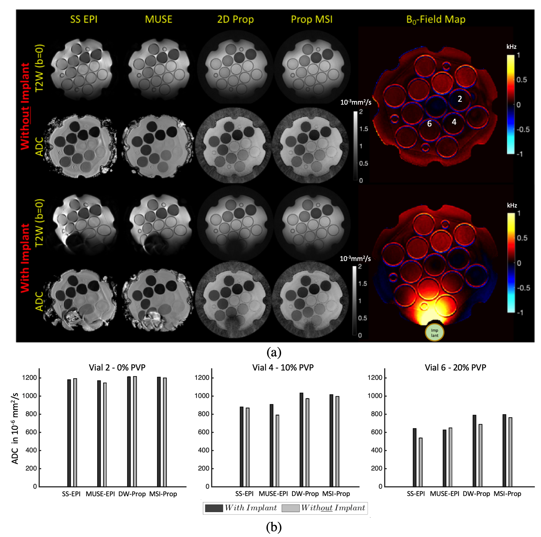
Fig.3. (a) Representative T2W(b=0) images and associated ADC maps: ADC's are shown for each of the four evaluated methods. Significant distortion seen in the SS-EPI, both with and without the presence of the metal high susceptibility source. Without the metal, image distortions were relatively mitigated with MUSE EPI. Qualitatively, DW-Prop and MSI-Prop were less susceptible to susceptibility artifacts. Vial 6 was largely non-viable in the SS-EPI and MUSE-EPI, but showed perfect geometry in DW & MSI-Prop. Vial 6 were compromised in DW-Prop, but were fully recovered in MSI-Prop.
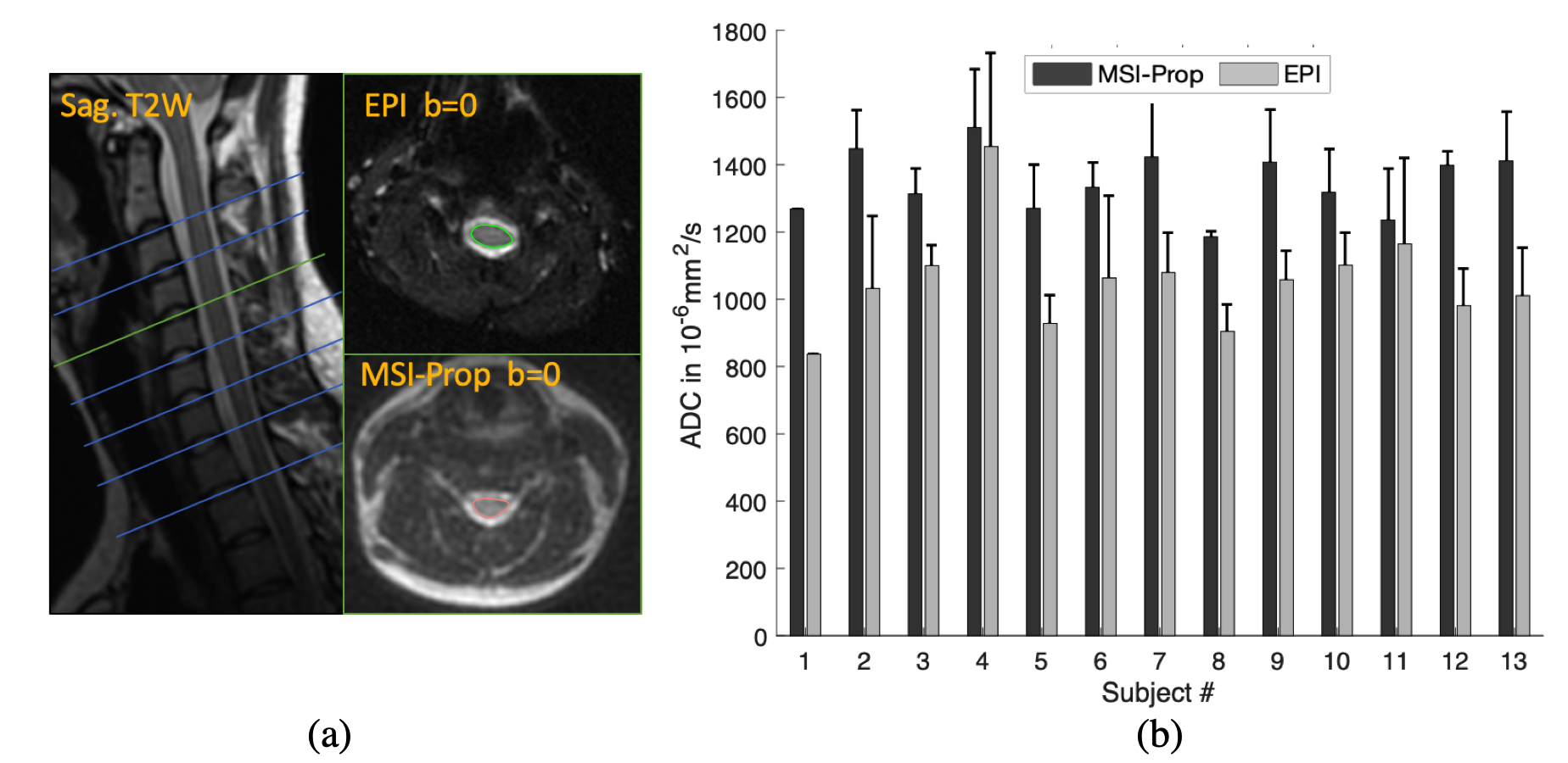
Fig.4. (a) Representative EPI and MSI-Prop T2W (b=0) images and spinal cord ROIs. Significant distortions can be seen for the EPI. MSI-Prop provided more anatomically accurate imaging of the cord. (b) ADC values computed with MSI-Prop were roughly 20% larger on average than the FOCUS EPI images. The computed bias was 293·10-6mm2/s among 13 subjects’ mean cord ADC values.