Arun Venkataraman1, Benjamin Risk2, Deqian Qiu3,4, Jianhui Zhong1,5, Feng (Vankee) Lin6,7, and Zhengwu Zhang8
1Physics and Astronomy, University of Rochester, Rochester, NY, United States, 2Biostatistics and Bioinformatics, Emory University, Atlanta, GA, United States, 3Radiology and Imaging Sciences, Emory University School of Medicine, Atlanta, GA, United States, 4Biomedical Engineering, Emory University, Atlanta, GA, United States, 5Imaging Sciences, University of Rochester Medical Center, Rochester, NY, United States, 6Brain and Cognitive Sciences, University of Rochester, Rochester, NY, United States, 7Neuroscience, University of Rochester Medical Center, Rochester, NY, United States, 8Biostatistics and Computational Biology, University of Rochester Medical Center, Rochester, NY, United States
1Physics and Astronomy, University of Rochester, Rochester, NY, United States, 2Biostatistics and Bioinformatics, Emory University, Atlanta, GA, United States, 3Radiology and Imaging Sciences, Emory University School of Medicine, Atlanta, GA, United States, 4Biomedical Engineering, Emory University, Atlanta, GA, United States, 5Imaging Sciences, University of Rochester Medical Center, Rochester, NY, United States, 6Brain and Cognitive Sciences, University of Rochester, Rochester, NY, United States, 7Neuroscience, University of Rochester Medical Center, Rochester, NY, United States, 8Biostatistics and Computational Biology, University of Rochester Medical Center, Rochester, NY, United States
In this study, we quantified the effects of simultaneous multislice and phase acceleration on dMRI data quality, and how patient factors such as motion influenced data quality. Faster sequences showed less patient motion, but had a drop in signal-to-noise ratio and contrast-to-noise ratio.
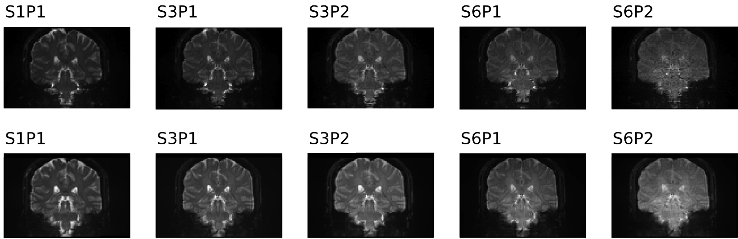
Figure 2 Representative section from Figure 1 normalized to highest intensity of each acquisition. The noise amplification in the midbrain and brain stem is easily visualized here (right-most sections in each row).
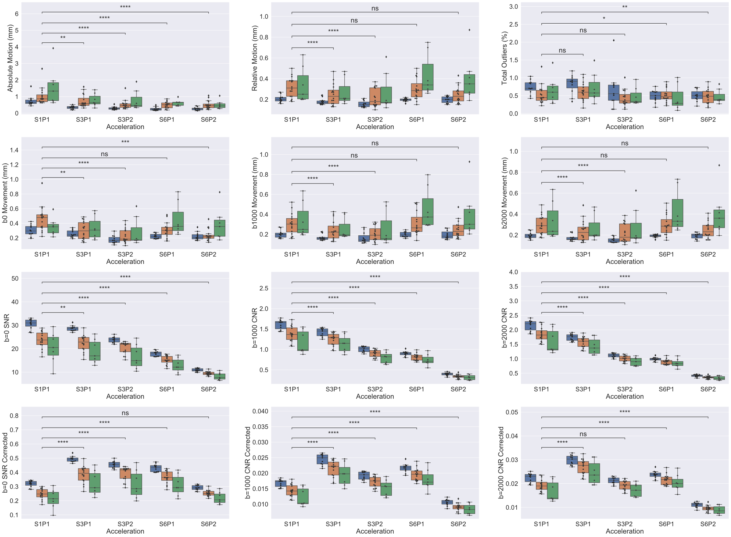
Figure 3 Boxplots representing quality control metrics (described on y-axis labels) over different acceleration schemes (x-axis). Blue bars indicate young subjects, orange indicates healthy, old subjects, and green indicates MCI subjects. p-values calculated using a Generalized Estimating Equation (GEE) with Gaussian assumption and exchangeable correlation accounting for repeated subject scans. p-values reported after false discovery rate (FDR) correction; significance: *p<0.05, **p<0.01, ***p<0.001, ****p<0.0001.