Malte Hoffmann1,2, Daniel C Moyer3, Lawrence Zhang3, Polina Golland3, Borjan Gagoski1,4, P Ellen Grant1,4, and André JW van der Kouwe1,2
1Department of Radiology, Harvard Medical School, Boston, MA, United States, 2Department of Radiology, Massachusetts General Hospital, Boston, MA, United States, 3Computer Science and Artificial Intelligence Laboratory, MIT, Cambridge, MA, United States, 4Fetal-Neonatal Neuroimaging and Developmental Science Center, Boston Children's Hospital, Boston, MA, United States
1Department of Radiology, Harvard Medical School, Boston, MA, United States, 2Department of Radiology, Massachusetts General Hospital, Boston, MA, United States, 3Computer Science and Artificial Intelligence Laboratory, MIT, Cambridge, MA, United States, 4Fetal-Neonatal Neuroimaging and Developmental Science Center, Boston Children's Hospital, Boston, MA, United States
Acquiring standard sagittal, coronal and axial planes with fetal brain MRI is challenging as frequent pose changes result in arbitrary orientations. We present a machine learning driven system that automatically prescribes standard orthogonal planes and demonstrate its utility in-vivo.
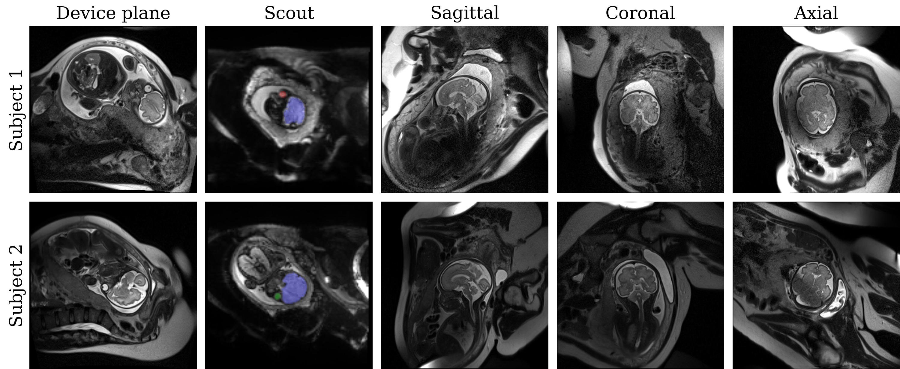
Figure 5. In-vivo evaluation in fetuses at 32 and 31 weeks' gestation. The fetal brain is arbitrarily oriented relative to the planes of the MRI scanner. The network detects the brain, left (red) and right (green) eyes from the full-uterus scout, oriented along the device axes (sagittal to the mother). From these landmarks, we derive the head pose. The target sequence automatically acquires standard sagittal, coronal and axial views of the brain. We repeat the scout before each anatomical acquisition. The second fetus required several attempts due to substantial subject motion.
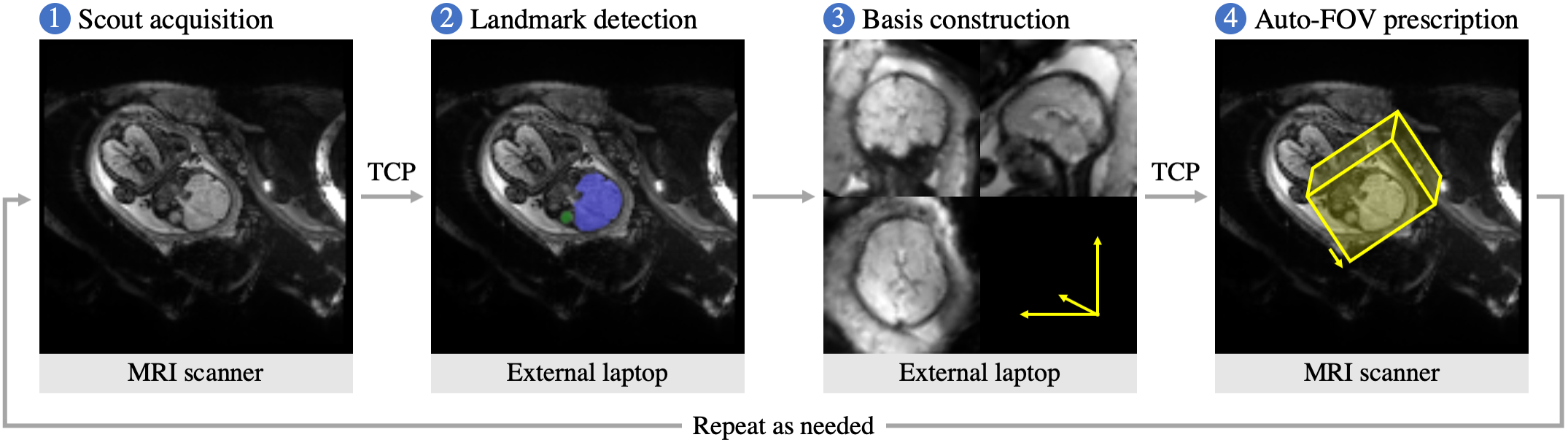
Figure 2. Automatic field-of-view prescription. First, the operator acquires a rapid full-uterus scout. Second, an external laptop receives the scout via TCP and hosts the network that detects the fetal brain and eyes. Third, we construct an anatomical basis from these landmarks. Finally, the target sequence receives the anatomical frame and acquires sagittal, coronal or axial slices of the brain according to the operator’s selection. All communications are automatic, and the procedure can be repeated as needed to acquire different views or to respond to fetal head-pose changes.