Stefan Wampl1, Tito Körner1, Martin Meyerspeer1, Marcos Wolf1, Maxim Zaitsev1, and Albrecht Ingo Schmid1
1High Field MR Center, Center for Medical Physics and Biomedical Engineering, Medical University of Vienna, Vienna, Austria
1High Field MR Center, Center for Medical Physics and Biomedical Engineering, Medical University of Vienna, Vienna, Austria
The capabilities of established computer vision object tracking algorithms on MR images is demonstrated. The image-based object trackers available from OpenCV are a perfect fit for retrospective and prospective motion correction methods.
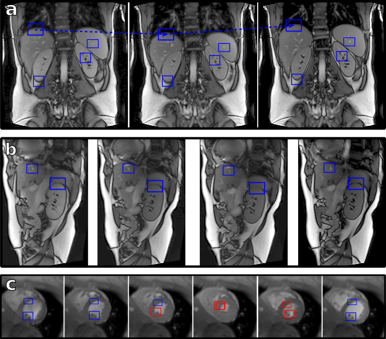
Object tracking on (a) a coronal and (b) a parasagittal image series of the abdomen and thoracal borders (3T, TrueFISP, FOV 384x384 mm2, 256x256 matrix, TR/TE 360/1.46) with bounding boxes independently tracking different landmarks on the images (diaphragm, liver, spleen, kidney). (c) Object tracking performed on a CINE acquisition of the heart. The target is missed during end-systole (red boxes) when the image features change, but reestablishes the position back in diastole.
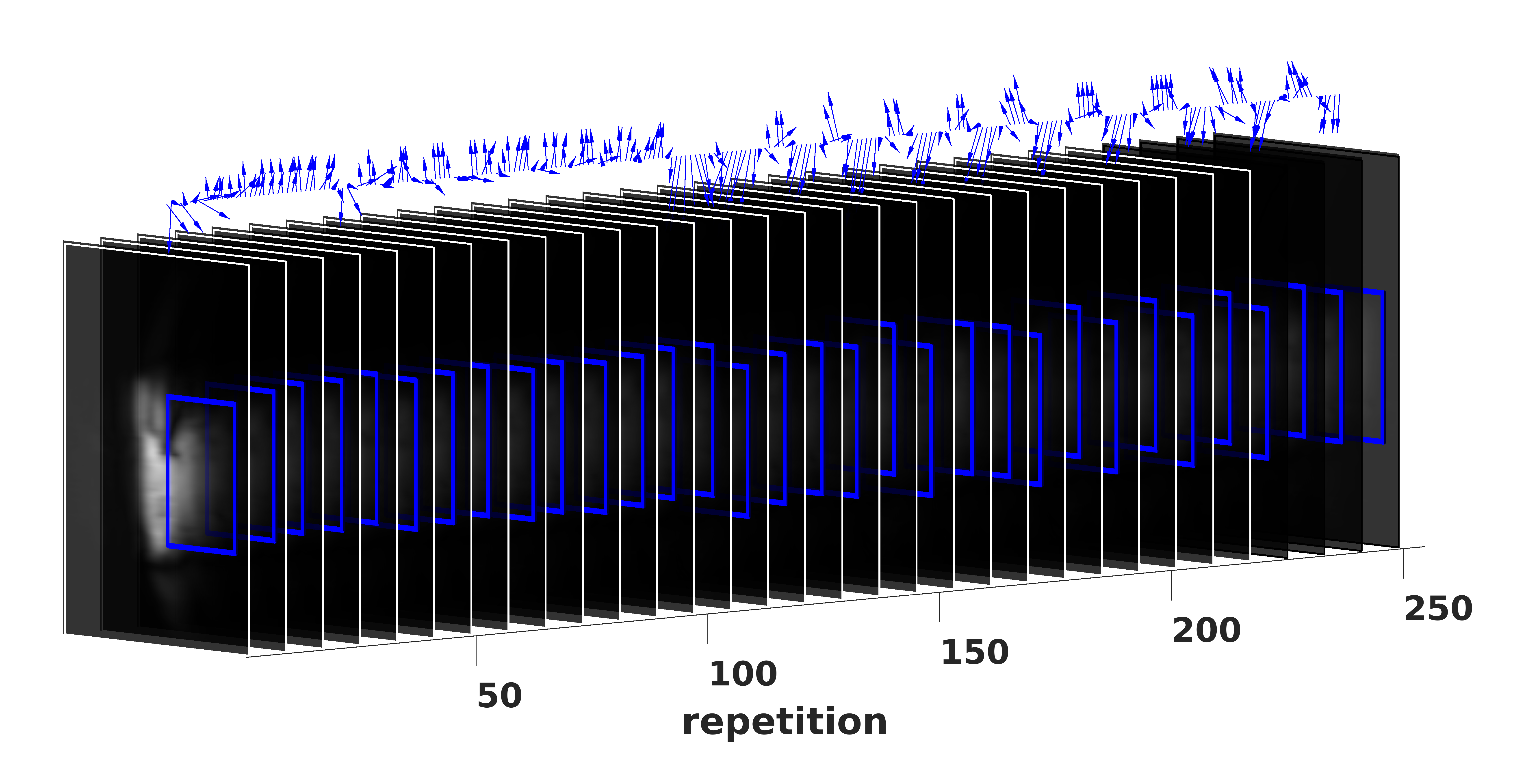
Tracking of the heart on a series of fast low-resolution image navigators (sagittal, 256 repetitions, every 8th displayed) as appropriate for prospective motion correction of cardiac MR spectroscopy. The bounding box follows the heart well during several respiratory cycles, with heavy breathing starting from repetition 100. The detected in-plane motion, as indicated by the series of arrows on top, is exaggerated by a factor of 3 for visualization purposes.