Filiz Yetisir1, Esra Abaci Turk1,2, P. Ellen Grant1,2,3, Elfar Adalsteinsson4,5, and Lawrence L. Wald3,5,6
1Fetal-Neonatal Neuroimaging and Developmental Science Center, Boston Children's Hospital, Boston, MA, United States, 2Department of Pediatrics, Harvard Medical School, Boston, MA, United States, 3Department of Radiology, Harvard Medical School, Boston, MA, United States, 4Department of Electrical Engineering and Computer Science, Massachusetts Institute of Technology, Cambridge, MA, United States, 5Harvard-MIT Division of Health Science and Technology, Massachusetts Institute of Technology, Cambridge, MA, United States, 6Athinoula A. Martinos Center for Biomedical Imaging, Massachusetts General Hospital, Charlestown, MA, United States
1Fetal-Neonatal Neuroimaging and Developmental Science Center, Boston Children's Hospital, Boston, MA, United States, 2Department of Pediatrics, Harvard Medical School, Boston, MA, United States, 3Department of Radiology, Harvard Medical School, Boston, MA, United States, 4Department of Electrical Engineering and Computer Science, Massachusetts Institute of Technology, Cambridge, MA, United States, 5Harvard-MIT Division of Health Science and Technology, Massachusetts Institute of Technology, Cambridge, MA, United States, 6Athinoula A. Martinos Center for Biomedical Imaging, Massachusetts General Hospital, Charlestown, MA, United States
Two-channel
RF shimming at 3T can be used to improve transmit field amplitude and uniformity
for fetal MRI without increasing maternal or fetal SAR in some pregnant
subjects. The biggest difference in imaging performance and SAR patterns is observed between left lateral and supine models.
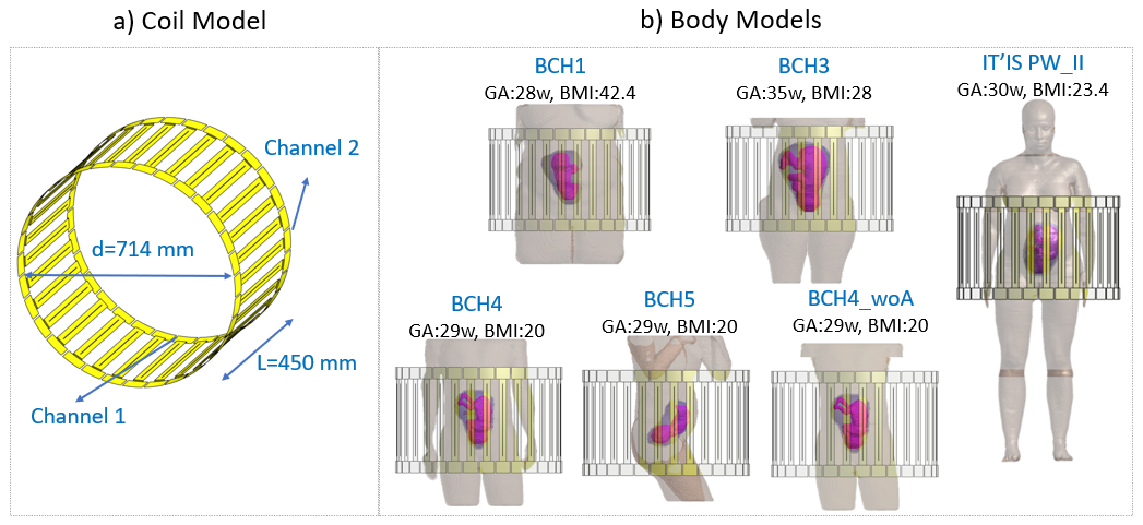
Figure 1: Numerical model of the
2-channel, 32-rung high pass birdcage coil (a) and the numerical pregnant body
models (b) used in this study. Only the skin, uterus and fetus are shown in the
body models for simplicity. GA: gestational age, w: weeks, BMI: body mass
index.
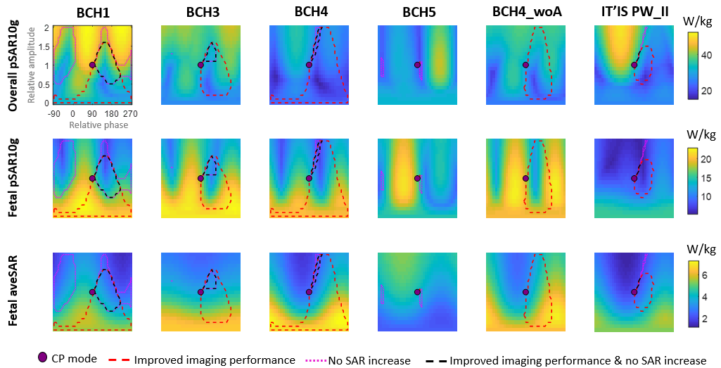
Figure 3: Overall (maternal and
fetal) peak local SAR, fetal peak local SAR and fetal average SAR for different RF shim
settings where relative amplitude and phase of the two channels is varied from 0 to 2 (vertical axis) and from -90° to 270° (horizontal axis) respectively. All shim settings are normalized to maternal
whole-body average SAR of 2 W/kg. CP mode: circularly polarized birdcage mode,
improved imaging performance: both average B1+ and B1+ variation
is improved compared to CP mode, no SAR increase: maternal or fetal SAR does
not increase compared to CP mode.