Sriranga Kashyap1, Roy A. M. Haast2, Thomas F. Kirk3, An T. Vu4,5, Denizhan Kurban1, Ron Hellenbrand1, Christopher J. Wiggins6, Alard Roebroeck1, Ali R. Khan2, David A. Feinberg1,7,8, Benedikt A. Poser1, and Dimo Ivanov1
1Department of Cognitive Neuroscience, Maastricht University, Maastricht, Netherlands, 2Centre for Functional and Metabolic Mapping, Western University, London, ON, Canada, 3University of Oxford, Oxford, United Kingdom, 4University of California, San Francisco, CA, United States, 5San Francisco Veteran Affairs Health Care System, San Francisco, CA, United States, 6Scannexus B.V., Maastricht, Netherlands, 7Advanced MRI Technologies, Sebastopol, CA, United States, 8Helen Wills Neuroscience Institute, University of California, Berkeley, CA, United States
1Department of Cognitive Neuroscience, Maastricht University, Maastricht, Netherlands, 2Centre for Functional and Metabolic Mapping, Western University, London, ON, Canada, 3University of Oxford, Oxford, United Kingdom, 4University of California, San Francisco, CA, United States, 5San Francisco Veteran Affairs Health Care System, San Francisco, CA, United States, 6Scannexus B.V., Maastricht, Netherlands, 7Advanced MRI Technologies, Sebastopol, CA, United States, 8Helen Wills Neuroscience Institute, University of California, Berkeley, CA, United States
In this 7T study, we show that B1+ distribution can be improved by optimising the placement of dielectric pads. We demonstrate that B1+ has a direct impact on perfusion measurements using ASL at 7T and therefore, on potential clinical utility.
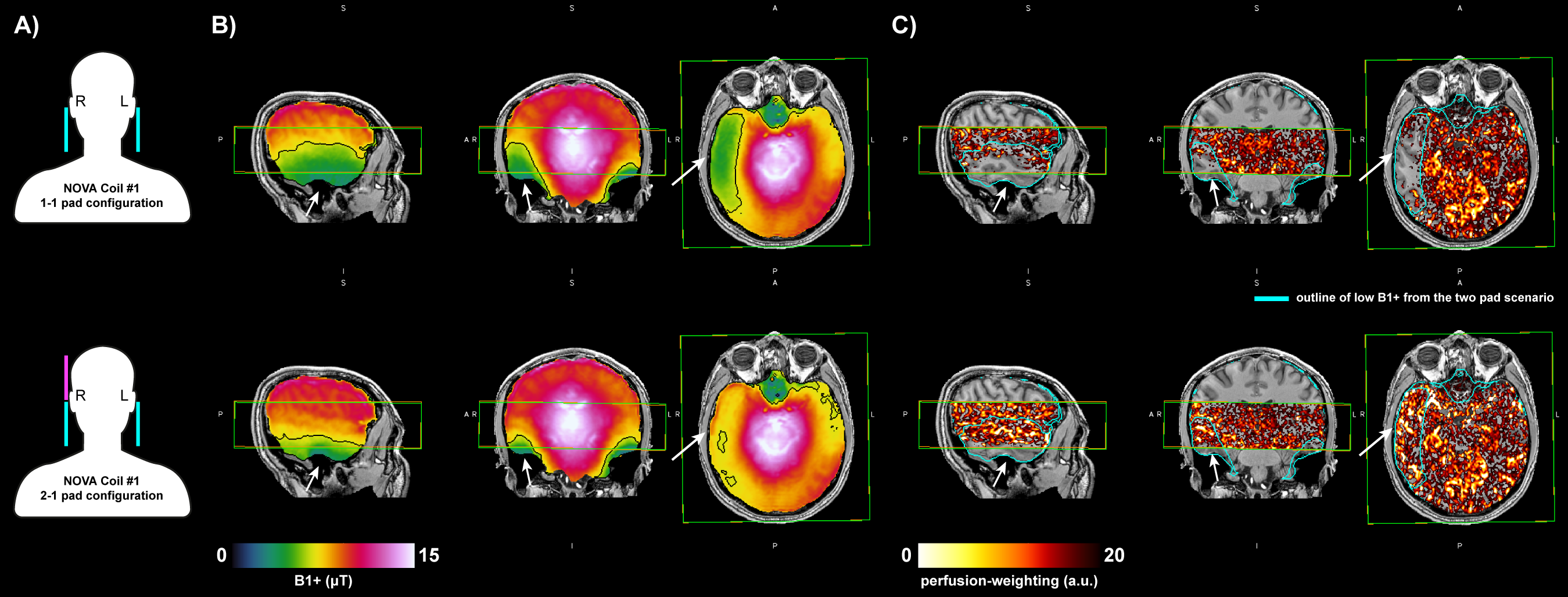
Figure 3: The data acquired on subj-02 on the same scanner using NOVA Coil #1 with two different dielectric pad configurations. (A) Schematic of dielectric pads placement (pink is the additional pad used), (B) B1+ maps (C) perfusion-weighted images. Overlaid rectangular outline indicates the slab coverage for perfusion imaging and cyan outline is the low B1+ from the 1-1 configuration. Note the impact of the 2-1 pad configuration, best illustrated in the axial slices.
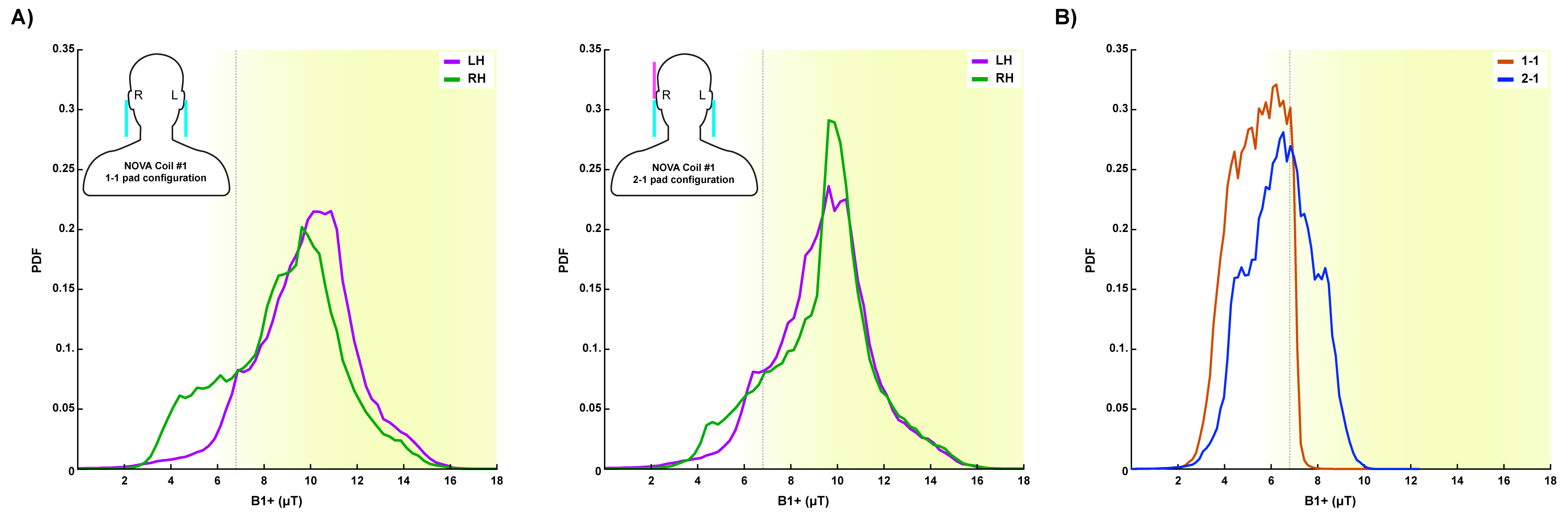
Figure 4: (A) B1 histograms in left and right hemispheres of subj-02 for 1-1 and 2-1 dielectric pad configuration (inset schematic) (B) Histogram showing the improvement in B1+ in the 2-1 configuration (region-of-interest is the low B1+ outline in Fig 3B top row). Yellow background and dotted line indicate 95% efficiency threshold for the tr-FOCI inversion pulse.